
By Coleman Harris
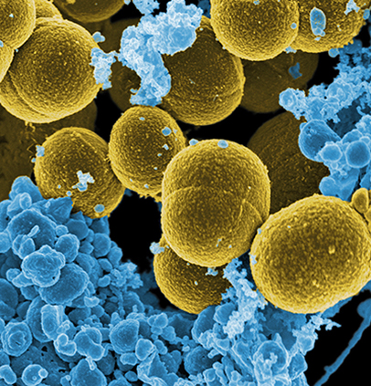
Staphylococcus aureus (S. aureus) is a pathogen that often causes the typical “staph infections” that form abscesses or boils. The pathogen maximizes its ability to infect a host via metabolites called siderophores, which acquire iron molecules to promote the growth and expansion of S. aureus. Iron is vital to bacteria like S. aureus – recognizing how they acquire iron can further the understanding of host-microbe interactions, and how best to stop them.
William Perry, a Chemistry graduate student working with the labs of Eric Skaar (Pathology, Microbiology and Immunology) and Richard Caprioli (Biochemistry), led research published in the Proceedings of the National Academies of Science that further explored the pathogen’s affinity for iron molecules. Specifically, they revisited the widely held assumption that S. aureus is starved of iron during colonization by mapping the distribution of the two siderophores involved in infection – staphyloferrin A (SA) and staphyloferrin B (SB).
Siderophores are thought to lead to the uniform uptake of iron, thus producing structurally homogeneous abscesses. However, emerging research suggests that some abscesses, like those formed by staph, exhibit heterogeneity in molecular structure and underlying differences in gene expression. Further, the advanced imaging methods used in this field suggest heterogeneity in molecular structure both among tissue types and within lesions. Thus, the researchers designed experiments to better understand the role of siderophores.
The researchers conducted two experiments in mice infected with S. aureus. The first experiment used high-performance MALDI IMS (matrix-assisted laser desorption/ionization imaging mass spectrometry), which is a method used to record the spatial distribution of molecules in tissue samples. In the researchers’ case, MALDI IMS was used to map the spatial distribution of SA and SB in the infected tissues.
The second experiment involved multiple integrated methods to visualize iron uptake in heart, kidney, and liver lesions – IMS (imaging mass spectrometry), histological staining to make structures in the cells visible, and fluorescence microscopy. The researchers utilized these to contrast the distributions of siderophores and iron molecules for different abscesses in the relevant tissues.
In the first experiment, SA had increased prevalence at most infection sites, while some sites had higher relative abundance of SB, suggesting that differences in iron uptake vary by site. This reinforces the researchers’ assertion that the two siderophores perform specific, heterogeneous roles, rather than influence pathogenesis homogeneously. And in the liver, heart, and kidney lesions, differential iron distributions were found across the S. aureus abscesses, again indicating differences in siderophore production and subsequent molecular response.
A key insight from this research is a better understanding of how the S. aureus pathogen acquires iron during infections via siderophores. While there is an understanding that siderophores have a role in the development of these infections, the results suggest that bacteria utilize different types for specific roles. Notably, the presence of SA at most infection sites suggests that it is important in the development of S. aureus abscesses, while the higher abundance of SB at some of the sites suggests its role in pathogenesis may be context dependent.
The study further illustrates the importance of technologies like MALDI IMS, along with the integration of multiple cellular imaging techniques, to image bacteria and understand molecular distributions. While clearly useful in the infection biology community, these techniques have further applications in producing cellular imaging data for the broader fields of microbiology and chemistry.