
By Sarah Glass
Scientists once dismissed the small, membrane-bound particles packed with proteins, nucleic acids, and lipids that are expelled by cells as mere cellular debris. In recent years, however, researchers have discovered that the particles, known as exosomes, serve as chemical signaling packets and play a vital role in intercellular communication. In a paper recently published in Nature Communications, Professor of Cell and Developmental Biology Alissa Weaver and her lab report a novel technique for visualizing exosomes in living organisms and detail new insights about exosome-mediated cellular migration, an activity central to such processes as embryonic development, wound healing, and the spread of cancer.
In the paper, first author Bong Hwan Sung and colleagues describe an improved technique for visualizing the secretion of exosomes. To more easily observe natural phenomena, researchers often attach a visual aid to a protein, such as a fluorescent tag, creating a reporter or a visual proxy that can be analyzed qualitatively and quantitatively.
Sung and team used a pH-sensitive fluorescent tag called pHluorin, modified to increase stability and brightness, and connected it to CD63, a molecule known to be present on exosomes. The resulting reporter construct, pHluo_M153R_CD63, is stably expressed in cells, marks exosomes released during live-cell imaging, and fluoresces green only in neutral conditions. Since cells live in neutral pH conditions but contain compartments that are highly acidic, the reporter fluoresces only when exosomes are secreted from the cell into the surrounding environment.
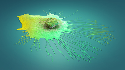
Among cells expressing pHluo_M153R_CD63, live imaging showed that exosomes are released at the front of the migrating cell, creating an exosomal trail for the next cell to follow. Using their novel reporter, the researchers visualized exosomes both in live cells and in live chicken embryos and mice, confirming the utility of their reporter in cultured cells as well as in whole organisms.
Finally, researchers added a second reporter to their construct that fluoresces red in acidic conditions, allowing for visualization of exosomes within the cell membrane. Using this dual reporter, the team discovered that after migrating cells detect an exosome trail, they engulf the exosomes, although the timeline varies significantly between cells. Overall, this work used a reporter system to enhance the visualization of cellular communication mediated by exosomes, giving the field a better look at how migration dynamics and cooperativity occur.
This research was supported by the National Institutes of Health and Vanderbilt University.