
A Cut Above

Present a surgeon with a challenging dilemma, and wheels begin turning. By their nature, surgeons are intuitive, adaptive problem-solvers. Think of those children who repaired their own broken toys, who constantly questioned conventional wisdom, and who thought so far outside the box, the box’s walls ceased to exist.
Driving these successes is innovation. Among Vanderbilt University Medical Center’s more than 240 clinical and research faculty, 118 residents and 19 fellows who make up the eight departments and 10 divisions of the Section of Surgical Sciences are brilliant minds in every specialty who imagine, innovate and invent, motivated by a dogged desire to improve patient care and advance surgical practice.
“The environment here allows that to happen because you’ve got learners who are excited and ask questions,” said Carmen Solorzano, MD, FACS, John L. Sawyers Professor of Surgery and chair of the Department of Surgery. “You have the teachers who are constantly looking for ways to improve how we take care of patients. And we are in proximity of all these smart researchers. Collaboration with others at the Medical Center and with the Medical School — with bioengineering, with engineering, with biophotonics — happens continually, just under the surface. And it’s a beautiful thing.”
All the essential ingredients have been purposefully put into place to encourage exploration of new ideas, said Seth Karp, MD, H. William Scott Jr. Professor, director of the Vanderbilt Transplant Center and chair of the Section of Surgical Sciences.
“This is the most important thing we try to create: a field for innovation,” he said. “We recruit people who want to be innovative; we set up programs that support innovation, and we build collaborations that support new ways of thinking. We fund innovation, and we spend a significant amount of our budget on new technologies. We never actively discourage anyone from pursuing an idea. If we feel like the idea is not perfect, we try to perfect it.”
Robot-assisted hernia repair means shorter hospital stays, fewer complications
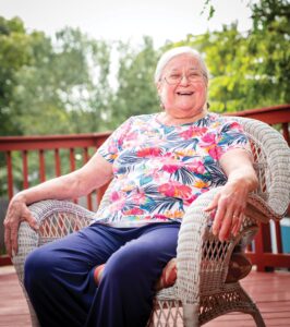
When Antioch resident Diane Ray felt a large bulge in her abdomen, she worried it might be cancer. Finding out it was a hernia was a huge relief.
Because she had experienced a tough recovery from a previous emergency gallstone surgery with a large, open incision, learning the hernia repair could be made using a minimally invasive, robot-assisted procedure reduced her anxiety even more.
“After they open up your belly and use 15 staples to close it, you definitely want to hear that there is a less-invasive way to get a repair done,” Ray said. “I may be 75, but when Dr. Bradley told me he could use a robot in a minimally invasive surgery, I was all for it.”
Ray’s surgeon, Joel Bradley III, MD, assistant professor of Surgery, is a member of the Vanderbilt Hernia Center, a regional center for hernia repairs. Surgical faculty have specialized training and experience to perform minimally invasive and robot-assisted hernia repair for the most complex cases, including patients with multiple and occult (hidden) hernias.
Vanderbilt University Medical Center is among just a few medical providers in Tennessee offering a robot-assisted, enhanced-view, totally extraperitoneal (eTEP) hernia repair, a minimally invasive procedure that replicates a hernia repair performed with a large, open incision. Vanderbilt Hernia Center surgeons also train other surgeons throughout the U.S. on the eTEP procedure.
“The use of robotics in abdominal wall reconstruction or hernia surgery has really transformed our approach to comprehensive hernia care,” said Joseph Broucek, MD, who serves as co-director of the Vanderbilt Hernia Center, along with Richard Pierce, MD, PhD, FACS. “The ability to recreate what we used to do through a big open abdominal incision using just three or four small incisions that are less than a centimeter has totally changed the game.
“Also, the amount of pain a patient experiences and the time they spend in the hospital have both decreased significantly. The vast majority of patients barely require any narcotic after the recovery room and are happy on just acetaminophen and ibuprofen going home.”
At VUMC, the length of stay for an open Rives-Stoppa ventral hernia repair is three days, but with an eTEP repair, the identical procedure using robotics, that’s been reduced to 1.09 days. The rate of surgical site occurrences — such as an infection or a buildup of fluids — has been reduced by nearly half in the eTEP group compared to the rate of these complications in the open surgery group.
Using robotics also gives the surgeon greater precision to place supportive mesh through small incisions, including between the abdominal muscle and the posterior rectus sheath (fascia) or between the muscle and the peritoneum (the abdominal lining).
“With robotics, we’re able to stay out of the abdominal cavity entirely and just work within the abdominal wall to repair the muscle,” Bradley said. “This reduces the amount of fixation needed to hold it in place, because the mesh is sandwiched within the muscles so it can’t really move anywhere.”
“My recovery time was remarkably easy, which to me is the best part of this,” Ray said. “I didn’t have to spend two or three nights in the hospital to make sure I was stable. One night was all it took. They gave me the usual postoperative medications for pain, but happily I didn’t have to take a single one.” – By Jill Clendening
Chemo-surgery-radiation combo brings hope for those with pancreatic cancer
Art Rich never gave up hope when he was diagnosed with pancreatic cancer in October 2018 and told it had grown too much for surgery to be an option. But he was frustrated. The diagnosis, which included three biopsies, had already taken about two months.
“The process seemed to be terribly slow,” he said. “Even though they said I might have cancer, there seemed to be no urgency.”
He wanted a second opinion, so he went to Vanderbilt-Ingram Cancer Center after a friend told him about Kamran Idrees, MD, MSCI, MMHC, Ingram Associate Professor of Cancer Research. Idrees, who is chief of the Division of Surgical Oncology and Endocrine Surgery, specializes in treating complex cancers of the pancreas, liver and bile ducts that might involve major blood vessels or adjacent organs.
“We are doing surgeries that are not being done at other major hospital systems in Nashville or in neighboring states,” said Idrees, who also serves as director of Pancreatic and Gastro-Intestinal Surgical Oncology.
One of those surgeries entails treating patients with chemotherapy before resection of the pancreas, an approach that benefits patients who may have been told their cancer is inoperable. (Typically, pancreatic surgeries are performed first with “operable” pancreatic cancers, i.e., those that don’t involve major blood vessels, followed by chemotherapy.)
“If pancreatic cancer patients who are deemed unresectable come to us for a second opinion, in a certain subset of patients, we are able to do the surgery,” Idrees said. “That’s where the value of a second opinion comes in.”
Idrees works closely with a multidisciplinary team of surgeons, medical oncologists, radiation oncologists, radiologists and pathologists to monitor the tumor’s response to the chemotherapy, checking to see when it shrinks enough to proceed with the surgery. The size of the tumor is but one concern. The tumor’s involvement with major blood vessels that supply major organs is a primary consideration. When Idrees performs the surgeries, he coordinates with vascular or organ transplant surgery teams who are highly experienced in reattaching major blood vessels.
However, when Rich first went to Idrees, he learned his case was a particularly challenging one. The surgeon initially told him the tumor appeared to be unresectable, but he promised to continue monitoring response to the chemotherapy. Dana Cardin, MD, MSCI, associate professor of Medicine in the Division of Hematology and Oncology, treated the cancer with rounds of an aggressive combination of multiple chemotherapies for six months, then referred him for radiation therapy.
He then went on a maintenance program with the chemotherapy capecitabine, taking the drug orally for two weeks, then one week off. For months, there seemed to be not enough response for Rich to undergo surgery.
But about a year and a half after having started the chemotherapy treatments, he asked Cardin if his prospects for surgery had changed. She scheduled another appointment for him with Idrees.
“Dr. Idrees came bounding into the examination room,” Rich said. “The very first thing I noticed is that his body language was radically different than in our previous meeting. He was much more positive and optimistic, and he walked us through what would happen on the surgical table.”
The tumor had finally responded to the point that it could be removed. Rich underwent a 14-hour surgery on Oct. 30, 2020. He is now cancer free.
“If you looked at me without knowing what I’ve gone through, you would never know,” Rich said. “People tell me I look healthier than I have ever looked. They are absolutely amazed.”
Vanderbilt University Medical Center is the only Pancreatic Cancer Center of Excellence as designated by The National Pancreas Foundation in Tennessee, Alabama, Mississippi or Arkansas.
Vanderbilt offers pancreatic cancer patients second-opinion evaluations, a dedicated nurse navigator to assist them, a multidisciplinary clinic and a protocol that expedites time to treatment. – By Tom Wilemon
3D modeling guides minimally invasive surgical removal of child’s tumor

Tekoa Morton was 3 years old when doctors found an inoperable mass surrounding multiple structures in her chest and abdomen. Her parents, Amanda and Colt Morton, chose to use alternative therapy to treat their daughter in hopes of avoiding the use of chemotherapies and radiation often prescribed in the treatment of cancer.
The tumor, stage 3 thoracoabdominal ganglioneuroblastoma, was attached to her aorta, renal vessels, ureter and diaphragm.
“For three years we used alternative therapies to monitor Tekoa, and things stayed relatively stable with some improvement,” said Amanda.
In summer 2020 Tekoa began complaining and crying about her stomach hurting. She was admitted to Monroe Carell Jr. Children’s Hospital at Vanderbilt, and scans showed that the mass had moved and was putting pressure on her nerve endings.
“We asked about partial resection possibilities because we still were not interested in chemo or radiation,” Colt said.
After days of examining images of Tekoa’s mass and anatomy, Irving J. Zamora, MD, MPH, assistant professor of Pediatric Surgery and director of Advanced Minimally Invasive Surgery at Children’s Hospital, met with the Mortons.
Six words started a new conversation: “I think I can get it,” Zamora said.
“We needed someone to come in with that level of confidence and empathy and connect with us,” said Amanda. “I remember just crying because we felt more at ease. He calmed us, and he was vested. It was such a relief.”
Zamora and his team used two innovative approaches to Tekoa’s care: minimally invasive surgery (MIS), the least intrusive pathway to performing a wide range of medical procedures and surgeries, combined with a unique 3D modeling innovation to assist in novel surgical approaches.
“We had just begun rebuilding our MIS program for tumors when Tekoa’s case was presented to us,” said Zamora. “The 3D model allowed us a unique opportunity to understand the complexity of the tumor and the relationship to surrounding structures.
“We were able to hold the model in our hands, study it and thoroughly examine it from every single angle and perspective to determine the best surgical approach,” he said. “While multiple imaging modalities are available, the use of 3D modeling took us to the next level.
“This allowed for precision-guided oncologic surgery using 3D modeling, and the outcome was fantastic.”
The recent addition of a state-of-the-art printer creates highly complex models, like in Tekoa’s case. The lifelike replica of her internal structure made all the difference, said Zamora.
MIS uses telescopes and operating instruments through small incisions. Surgeons at Children’s Hospital have extensive experience and expertise in MIS, which they have tapped into since the 1990s.
“We are building a program that is allowing us to do high-level, advanced surgical operations and meet the needs of some of the most critically ill, medically complex patients,” said Zamora.
Children’s Hospital offers MIS for a wide range of complex conditions with the goal of providing equal or superior clinical outcomes as well as reducing surgical risks, use of pain medications and hospital length of stay.
Soon after the completion of the surgery, five words confirmed the Mortons’ decision to move forward with the novel technology used to remove the tumor: “We got all of it,” Zamora said.
“He literally used three small incisions,” said Colt. “The tumor, which was twice the size of her kidney, was removed through her belly button. It was all done laparoscopically.”
Zamora and his team explained the surgery to Tekoa’s parents using the 3D model, and it made all the difference, the pair said.
“Our children are our lives. There was a huge sense of comfort because we could understand and see it,” said Amanda. “Dr. Zamora was able to walk us through what he was going to do. He knew what he was dealing with down to the nerve endings and vital organs.”
Today, Tekoa is vibrant, healthy, and after two sets of scans, cancer free.
“Looking at her today, you wouldn’t know she had surgery,” said Colt. “She looks so good, and she feels good. We give our Lord and Savior Jesus all the glory.
“We look at her and see hope, peace, thankfulness and gratitude. This technology changed our lives.” – By Jessica Pasley
Father, son benefit from jaw surgeries guided by 3D virtual planning
There is a growing number of surgical specialties at VUMC that have been revolutionized by the addition of 3D virtual surgical planning. Oral and maxillofacial surgeons now use the tool in 70% of their surgeries, whether it is correcting a jaw deformity, replacing a temporomandibular joint, or meticulously reconstructing a defect following a traumatic injury or ablative tumor surgery, estimates Sam McKenna, MD, DDS, chair of the Department of Oral and Maxillofacial Surgery.
Orthognathic surgery corrected an underbite and cross bite caused by a rogue basketball shot that injured Jeremy Kerr in middle school, initiating a long, painful journey for the now 49-year-old.
“After someone shot the ball, another player jumped first and got the rebound but elbowed me in the jaw as I was jumping up,” he said. “There was a loud pop, and everybody heard it. The next morning, my jaw was stuck halfway open.”
The incident marked the beginning of more than three decades of jaw popping, locking and chronic pain. The hoops injury likely contributed to one side of his lower jaw growing longer than the other, but genetics also played a role in his underbite and cross bite.
Ironically, it was Kerr’s son Tyler who struggled with an even more pronounced underbite and cross bite, who got his jaw malformations corrected first.
“My son and I were initially going to go through the surgeries together, but I chickened out at the last minute,” Kerr laughed. “After I saw the great result my son had, I decided to go ahead. I’m glad I did. I now have no popping, no clicking, no continually biting my jaw.”
Orthognathic surgery involves moving, lengthening or shortening the bones of the upper and lower jaw to correct alignment of the jaws and teeth and improve facial appearance.
Tyler’s surgical journey began with images of his skull taken with a Cone Beam CT scanner. Then, a vendor specializing in digital diagnostic and treatment imagery transformed the images into a 3D digital CT. McKenna could then clearly view how Tyler’s entire jaw was shaped and how it related to his bite.
“This technology that allows us to plan surgeries in a virtual platform has really revolutionized the management of these deformities,” McKenna said. “The basis for that planning is, of course, still our examination of the patient, assessment of the deformity, and making various measurements. But the three-dimensional CT allows us to sit at a computer screen and manipulate the bones in a very analytic and precise way.
“A surgical template based on the virtual planning is created using a 3D printer that we take to the operating room. That template allows us to achieve in the OR exactly what we’ve mapped out during the virtual planning.”
Prior to having this capability, McKenna and other Oral and Maxillofacial Surgery faculty and residents spent evenings and weekends in the lab making plaster models of patients’ jaws. The plaster was mounted onto a device called an articulator which allowed them to manually move the jaw models to plan surgeries.
“With the ability to do this virtually using the 3D CT scan, it really satisfied our long-standing desire to do this as analytically as possible, but with much greater accuracy,” McKenna said. – By Jill Clendening
Minimally invasive bariatric surgery puts officer back in the saddle
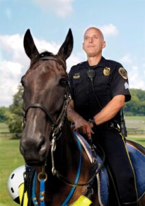
When Sgt. Kevin Hooper began supervising the Metro Nashville Park Police Mounted Patrol in 2013, he had one problem. The unit didn’t have a horse strong enough to safely support Hooper’s 419 pounds during long days on the job.
Fortunately for Hooper, Vanderbilt offers laparoscopic sleeve gastrectomy and laparoscopic gastric bypass, as well as revisional surgery for patients who have had previous, less successful bariatric procedures. Individuals with a body mass index (BMI) above 40 qualify, and individuals with a BMI of 35-40 can typically qualify if they have an obesity-related health issue such as sleep apnea, diabetes or heart disease. More than 99% of gastric procedures at VUMC, including revisional surgeries, are completed using a minimally invasive or laparoscopic approach, said Matthew Spann, MD, director of Vanderbilt Surgical Weight Loss.
Bariatric surgery, commonly called weight-loss surgery, not only reduces waistlines, but significantly lowers the risk of death related to many diseases including heart disease (40% lower), diabetes (92% lower), and cancer (60% lower), according to statistics from the American Society for Metabolic and Bariatric Surgery.
“It’s the reason I chose to specialize in bariatric surgery,” Spann said. “It’s rare that we can improve both quality of life and quantity of life with a single procedure; usually you can do one or the other. Seeing what happens for patients like Kevin makes our job exciting and rewarding.
“There is a greater than 90% chance that patients who are on one or two oral medications for diabetes will be able to come off those medications and stay off of them for at least five years. For patients with diabetes that requires insulin, there’s around a 50% chance they’ll be able to be insulin free and medication free five years after surgery, with a well-controlled hemoglobin A1c. And many folks will experience the benefits of better-managed diabetes before they ever leave a hospital. It’s a tangible and immediate impact.”
When a patient has a BMI greater than 35, their chance of reaching a normal body weight for a lasting period of time is less than 1%. In contrast, after bariatric surgery, around 80% of individuals will remain within 10-15% of their lowest weight, Spann said.
Unfortunately, only about 1% of people who qualify actually get surgery. That’s why the Vanderbilt Surgical Weight Loss team works to make bariatric surgeries more accessible. They meet with primary care providers throughout Tennessee to answer questions about obesity and the benefits of bariatric surgery, as well as share information on other tools available at Vanderbilt’s comprehensive weight loss centers.
Today, nearly four years after he underwent laparoscopic gastric bypass surgery at VUMC, Hooper has lost 200 pounds, and his partner as he patrols Metro Nashville’s 15,000-acre parks system is a jet-black Tennessee Walking Horse named Lex.
“I’m on a horse for work downtown almost every weekend now, and it wouldn’t be possible if I had not had the surgery,” Hooper said. “This totally changed the course of my future. And I was happy before, but with 100% certainty I can say I wouldn’t be nearly as happy if I wasn’t doing what I’m doing now for work.” – By Jill Clendening