
New technology helps pediatric patients who require frequent X-rays
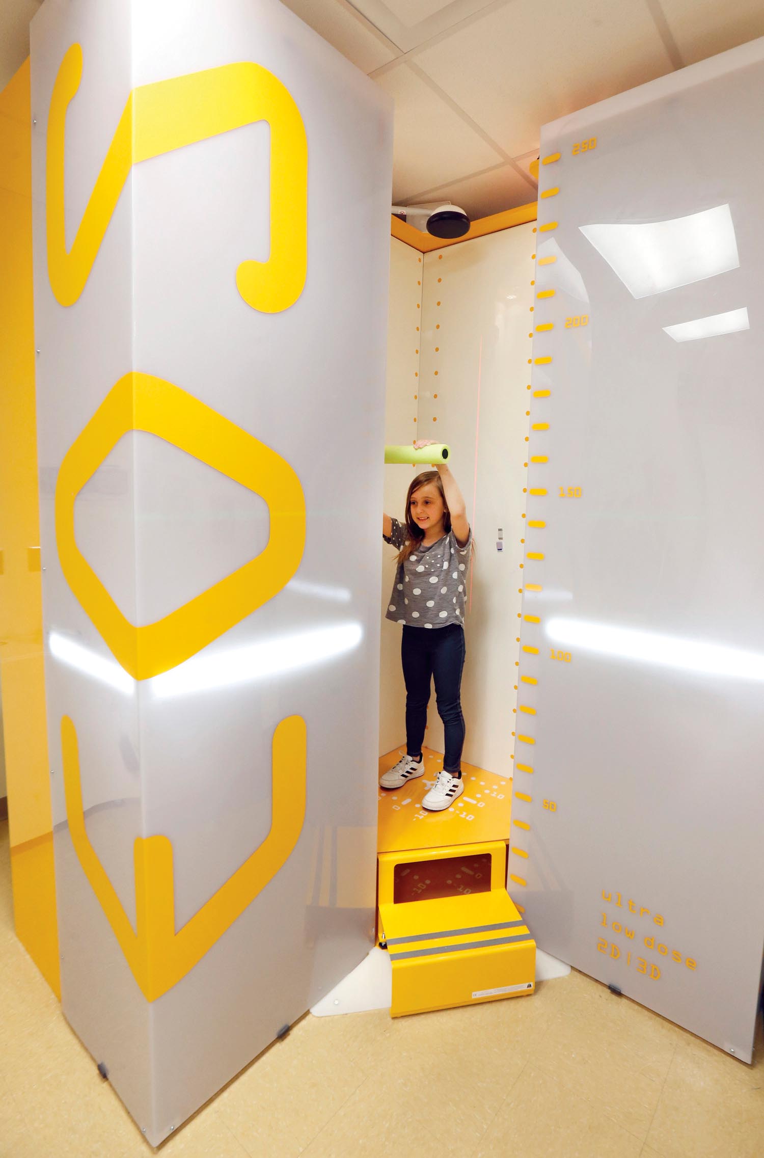
Chloie Jacobs, 9, prepares for a follow-up scan of her congenital scoliosis and climbs into a new X-ray imaging device at the pediatric orthopaedic clinic at Monroe Carell Jr. Children’s Hospital at Vanderbilt.
But this isn’t just any X-ray machine. For Chloie, the cutting-edge technology, known as EOS, feels more like a teletransporter, because of its outer space-like design. For her parents, Michael and Amber, the machine serves as peace of mind.
The EOS X-ray imaging system uses ultra-low radiation doses (up to 50 times lower depending on the scan type) to capture 2-D and 3-D images. The scan, complete in about eight to 15 seconds, obtains an image of the body in an upright, load-bearing position, which is more representative of the body’s natural function.
Previously, these patients had radiologic images taken with traditional X-ray machines and higher amounts of radiation, often in the supine position.
“She has X-rays at least three times a year, so anything with less radiation is always better,” Amber Jacobs said.
If a child can’t stand unassisted in the EOS machine, a chair specifically designed for the machine allows the child to sit while being scanned. Less radiation for high-quality scans is appealing to doctors and parents because the cumulative effects of too much radiation can be harmful to a child, increasing the risk of cancer.
“With children and adolescents, the goal is always to minimize the radiation dose. This is the ALARA concept: ‘as low as reasonably achievable.’ The EOS technology allows for substantial reduction in radiation exposure,” said Jeff Martus, MD, associate professor of Orthopaedic Surgery.