By Lorena Infante Lara
New technologies allow us to look at old knowledge with fresh eyes. Scientists have known about microvilli for a long time. Electron microscopy showed us how mature microvilli pack densely on the apical surface of gut, renal, and other epithelial cells, but it couldn’t show us how they form this structure. A recent Developmental Cell paper from the lab of Matt Tyska (Cell and Developmental Biology) explores the process of microvilli maturation – and reveals their previously unknown motility.
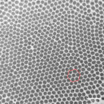
Microvilli are present on cells all over the body, helping to increase the surface area of cells to facilitate nutrient and solute absorption. Each cell can have upward of 3,000 microvilli on its surface, all neatly arranged in a honeycomb-like, hexagonal pattern. The work presented here comes as a follow up to a 2014 Cell paper by the Tyska lab that showed that mature microvilli physically attach to one another through adhesion complexes.
“The cells don’t grow all 3,000 microvilli exactly at the same time,” Tyska said. Microvilli grow stochastically all over the surface of a cell, so he reasoned that there must be a mechanism to bring them together and organized into the tight packing seen in the image.
Using lattice light-sheet microscopy and spinning disk confocal microscopy, Tyska’s lab monitored the growth of microvilli in live cells and discovered an unexpected dynamism in the packing process. Using actin – their primary structural element – as a motor, microvilli constantly glide across the cell surface.
“We didn’t know that microvilli moved at all,” Tyska said. “It’s a novel form of motility that we have discovered.”
The collisions between microvilli lead to the formation of adhesion complexes, such that, eventually, all microvilli pack so densely that they adopt the hexagonal structure, one of nature’s most efficient packing methods.
Vanderbilt’s lattice light-sheet microscope is one of only a few that exist in the world, custom built through the Biological Microscopy, Innovation, Immersion, and Discovery (BioMIID) program. BioMIID is a trans-institutional effort designed to help students, postdocs, and faculty from across campus innovate by building microscopy systems that do not yet exist in the commercial realm to answer previously unanswerable questions in biomedicine.
Tyska indicated that Vanderbilt will soon begin building a second lattice light-sheet microscope, and that the research community will be able to use it for their own projects.