Shane Watson, James Hayes, and Emma Koory are the winners of the inaugural Cell Imaging Shared Resource Life Is Beautiful Image Contest. They worked with Jenny Schafer, CISR managing director and research associate professor of cell and developmental biology, Oleg Kovtun, research assistant professor of chemistry, and CISR senior research specialists Kari Seedle and Tegy Vadakkan.
Located within the School of Medicine Basic Sciences, CISR is an institutional, fee-for-service advanced light and electron microscopy resource. The CISR provides researchers with access to state-of-the-art imaging equipment and expert technical support for sophisticated microscopy and analysis of tissue and cellular anatomy and physiology.
In collaboration with Basic Sciences, CISR, which has approximately 250 unique microscope users per year, wanted to showcase the beautiful images captured by users as part of their biomedical research at Vanderbilt. The annual CISR image contest was created to highlight these images.
“We are thrilled to have these three beautiful images shared by our CISR users! The CISR maintains instruments across numerous light and electron microscopy technologies, and the variety of samples that can be imaged on CISR instruments is evident from these winners,” Schafer said. “The stunning, whole mouse brain image acquired by Shane Watson shows the large end of the imaging spectrum, in which users can visualize connections across whole tissue or organs with light-sheet microscopy. The amazing image by James Hayes shows the smaller end of the light microscopy spectrum with super-resolution microscopy for imaging sub-cellular organelles. The beautiful image by Emma Koory also shows super-resolution microscopy with tremendous detail at the organelle level.”
First prize:
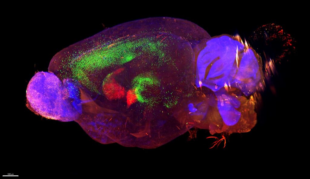
Watson’s research seeks to better understand the formation of synaptic inputs into the striatum, a region of the brain responsible for motor control, emotion, habit formation, and reward. He works with the protein latrophilin, a receptor known to mediate specific synaptic connections throughout the brain.
This image shows a whole mouse brain injected with two separate viruses: a Cre-dependent starter virus and a modified rabies virus. The starter virus infected Cre-expressing D2-type medium spiny neurons in the dorsal striatum (red).The modified rabies virus entered those same MSNs and traveled one synapse backward to denote retrograde, direct synaptic connections (green). This means that every green dot is an individual neuron that synapses directly onto the red, infected MSNs in the dorsal striatum.
Microscope: Z1 Zeiss Light Sheet Microscope
Objective lens: 2.5X
Microscopy technique: Fluorescence microscopy, light-sheet microscopy
Second prize:
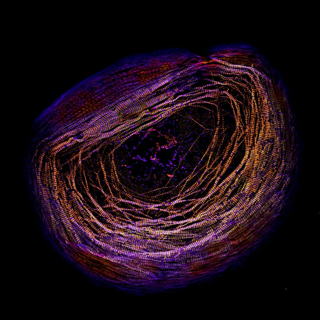
Hayes’ research focuses on how the beating muscle cells of the heart produce a contractile force. The human heart has thousands of cardiac myocyte cells like the one shown in the image to drive your heartbeat. The primary structures pictured in the cell are known as sarcomeres, which are the contractile units of muscle. The darker purple in the image is sarcomere actin filaments stained with phalloidin. The lighter gold and yellow colors are myosin filaments stained with an antibody. Myosin filaments pull on the sarcomere actin filaments to produce force, driving the heartbeat.
Microscope: Nikon CSU-W1 Spinning Disk Confocal with SoRa
Objective lens: 100X oil
Microscopy technique: Fluorescence microscopy, super-resolution microscopy
Third prize:
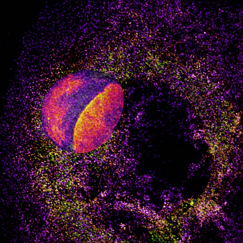
Koory studies cardiac development and injury and how the cells of the heart form sarcomeres, the structures that allow them to contract. The image shows a clefted nucleus amongst cellular mRNA, which was labeled with fluorescence in situ hybridization. This particular probe labels the polyA tail of all cellular mRNA. The nucleus was stained with DAPI. The mRNA channel was depth coded, resulting in multicolor dots. The nucleus was colored using the mpl-inferno LUT in Fiji.
Microscope: Nikon CSU-W1 Spinning Disk Confocal with SoRa
Objective lens: 100X (2.8X SoRa)
Microscopy technique: Fluorescence microscopy, super-resolution microscopy
Watson is a trainee in the lab of Richard Sando, assistant professor of pharmacology, and Hayes and Koory are trainees in the lab of Dylan Burnette, associate professor of cell and developmental biology.
Notably, all three winning images were all acquired from CISR instruments purchased through NIH S10 equipment grants. The Zeiss Z1 light-sheet microscope was awarded in 2019 through grant S10OD025109 and the Nikon Spinning Disk Confocal with SoRa was awarded in 2023 through grant 1S10MH130456.
Images were judged for scientific technique and aesthetic composition. Winners were selected from dozens of entries from staff and trainees in Basic Sciences, the School of Medicine, the School of Engineering, and Vanderbilt University Medical Center.