James Hayes, Rekha Nagarajan, and James Costanzo are the winners of the 2025 Cell Imaging Shared Resource Life Is Beautiful Image Contest. They worked with Jenny Schafer, CISR managing director and research associate professor of cell and developmental biology, Oleg Kovtun, research assistant professor of chemistry, and CISR senior research specialists Kari Seedle and Tegy Vadakkan to create these images as part of their individual research projects.
Located within the School of Medicine Basic Sciences, CISR is an institutional, fee-for-service advanced light and electron microscopy resource. CISR provides researchers with access to state-of-the-art imaging equipment and expert technical support for sophisticated microscopy and analysis of tissue and cellular anatomy and physiology.
With over 300 unique microscope users per year, CISR facilitates the creation of beautiful images captured by users as part of their biomedical research at Vanderbilt. The annual CISR image contest was created to highlight these images in collaboration with the SOMBS.
“We are ecstatic to see so many beautiful images submitted for our second year and thrilled to choose these three winners!” Schafer said.
Hayes was a Ph.D. student in the lab of Dylan Burnette, associate professor of cell and developmental biology, when he took the winning image. He is now a postdoctoral fellow in the lab of David Merryman, professor of pharmacology. Nagarajan, the second-place winner, is a Ph.D. student in the lab of Matt Tyska, the Cornelius Vanderbilt Professor of Cell and Developmental Biology at the School of Medicine Basic Sciences. Third-place winner Costanzo is a Ph.D. student in the lab of Vivian Gama, associate professor of cell and developmental biology and associate dean for mentoring. The images they submitted were taken with two of the CISR’s microscopes.
One of the microscopes, the Nikon CSU-W1 SoRa spinning disk confocal microscope, provides super-resolution imaging on a spinning disk confocal platform that allows increased spatial resolution and increased temporal resolution on the same system. Both the first- and third-place images were acquired on the Nikon SoRa. “The image by James Hayes of the zebrafish eye shows the power of high resolution in a large sample, while the image by James Costanzo shows the super resolution needed for imaging mitochondrial structure in individual cells,” Schafer said.
In turn, the Nikon AX R MP, a multiphoton microscope, allows for visualization that goes up to hundreds of microns deeper into samples compared to conventional confocal microscopes, which only allow limited depth imaging. “The image by Rekha Nagarajan is a fabulous example of the great depth achievable with the Nikon multiphoton, as it shows a whole-mouse small intestine with great length, width, and depth visualized in the full 3D volume.”
Submitted images were judged for scientific technique and aesthetic composition. Winners were selected from dozens of entries from staff and trainees in the School of Medicine Basic Sciences, the School of Medicine, Vanderbilt University Medical Center, and the School of Engineering.
Here are the winners.
First prize

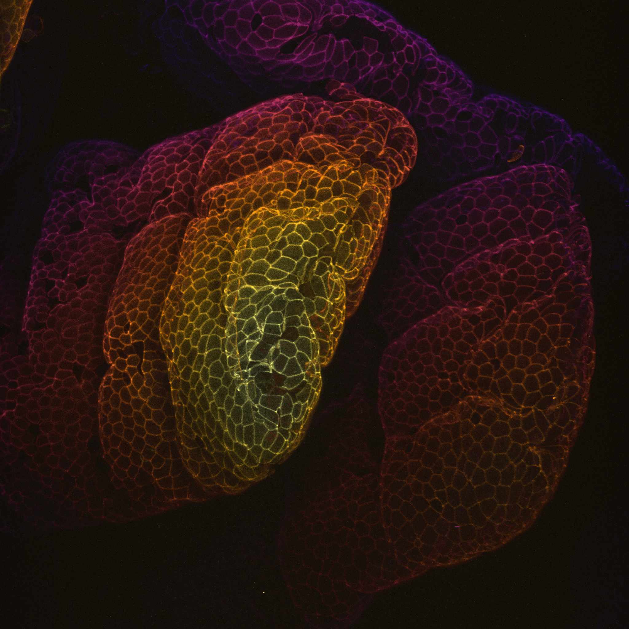
This zebrafish eye was labeled with a nuclear stain, which marks all cell nuclei, as well as a phalloidin stain, which marks the actin cytoskeleton.
Hayes’ doctoral research in the Burnette lab concerned proteins that regulate cardiovascular cell biology and contractility. “As part of that research, I frequently imaged zebrafish embryonic actin filaments,” Hayes said. “While the eye of the zebrafish embryo was not directly related to that research, it was always so striking under the oculars that I couldn’t resist capturing this image to share with other researchers or anyone interested in science!”
Microscope:
Nikon CSU-W1 SoRa spinning disk confocal microscope
Objective lens:
20X/0.8
Microscopy technique:
Fluorescence confocal
Second prize
Nonmuscle myosin-2C is a motor protein implicated in the shaping and bending of tissues, collective cell migration, and regulation of the paracellular permeability of epithelial sheets. It is seen here highly enriched at the junctional margins of the intestinal epithelial cells, where cell-cell contacts are formed. The endogenously tagged NM2C mouse allows us to fluorescently image the protein in its natural physiological context. The large protrusions captured here are called villi, tissue structures that line the walls of the small intestine to facilitate nutrient absorption and passage of fluids. Depth-coded coloring represents NM2C expression, with the deepest part of the villi shown in dark purple and the furthest point of the tip in whitish yellow.
Microscope:
Nikon AX R MP confocal microscope
Objective lens:
25X/1.1
Microscopy technique:
Fluorescence confocal, multiphoton confocal
Third prize
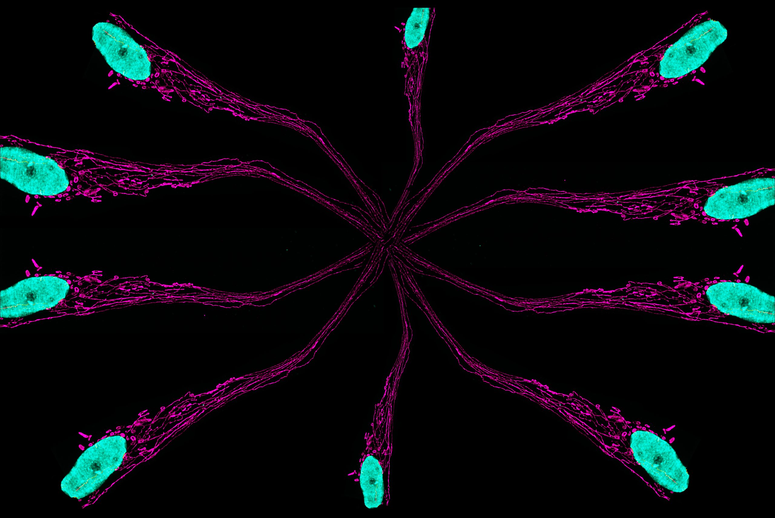
Immunofluorescence images were taken of induced pluripotent stem cell–derived, day 200+ neurons using Hoechst to stain for nuclei and anti-mitochondria antibodies to stain for mitochondria. The resulting image shows the mitochondria traveling through an axonal/dendritic projection from a single neuron. Deconvolution and duplication of the image resulted in a web-like design for an artistic effect.
Costanzo’s research explores how metabolic signaling influences cell fate transitions during neuronal differentiation. He uses patient-derived and isogenic iPSC models of EMPF1; encephalopathy due to defective mitochondrial and peroxisomal fission-1 is a rare neurodevelopmental disorder caused by mutations in dynamin-related protein 1 or DRP1. These mutations disrupt mitochondrial and peroxisomal fission, leading to abnormally elongated mitochondria and peroxisomes, as seen in both EMPF1 patients and iPSC-derived neural cells. Through this work, he is uncovering how changes in mitochondrial and peroxisomal structure influence neural development via their metabolic functions.
Microscope:
Nikon CSU-W1 SoRa spinning disk confocal microscope
Objective lens:
60X/1.42
Microscopy technique:
Confocal immunofluorescence microscopy
About the Cell Imaging Shared Resource
The Cell Imaging Shared Resource maintains instruments across numerous light and electron microscopy technologies and offers the latest technologies for Vanderbilt biomedical researchers. All three winning images were acquired using instruments that CISR purchased through NIH S10 equipment grants awarded in the last three years. The grant that allowed the purchase of the Nikon AX R MP confocal microscope (1S10OD032216-01A1) was awarded in 2022 and the grant to acquire the Nikon CSU-W1 SoRa spinning disk confocal microscope (1S10MH130456-01A1) was awarded in 2023.