Basic Sciences Featured Images
The following images have been generated by students, postdocs, staff, or faculty affiliated with Basic Sciences. Each one is the product of hard work, both technical and creative, and has been selected as the banner image for our newsletter, Basically Speaking. If you are interested in having your image featured in our newsletter and in this gallery, please submit it here.
For more images, please visit our Instagram page.
Evan Krystofiak – Cell Imaging Shared Resource
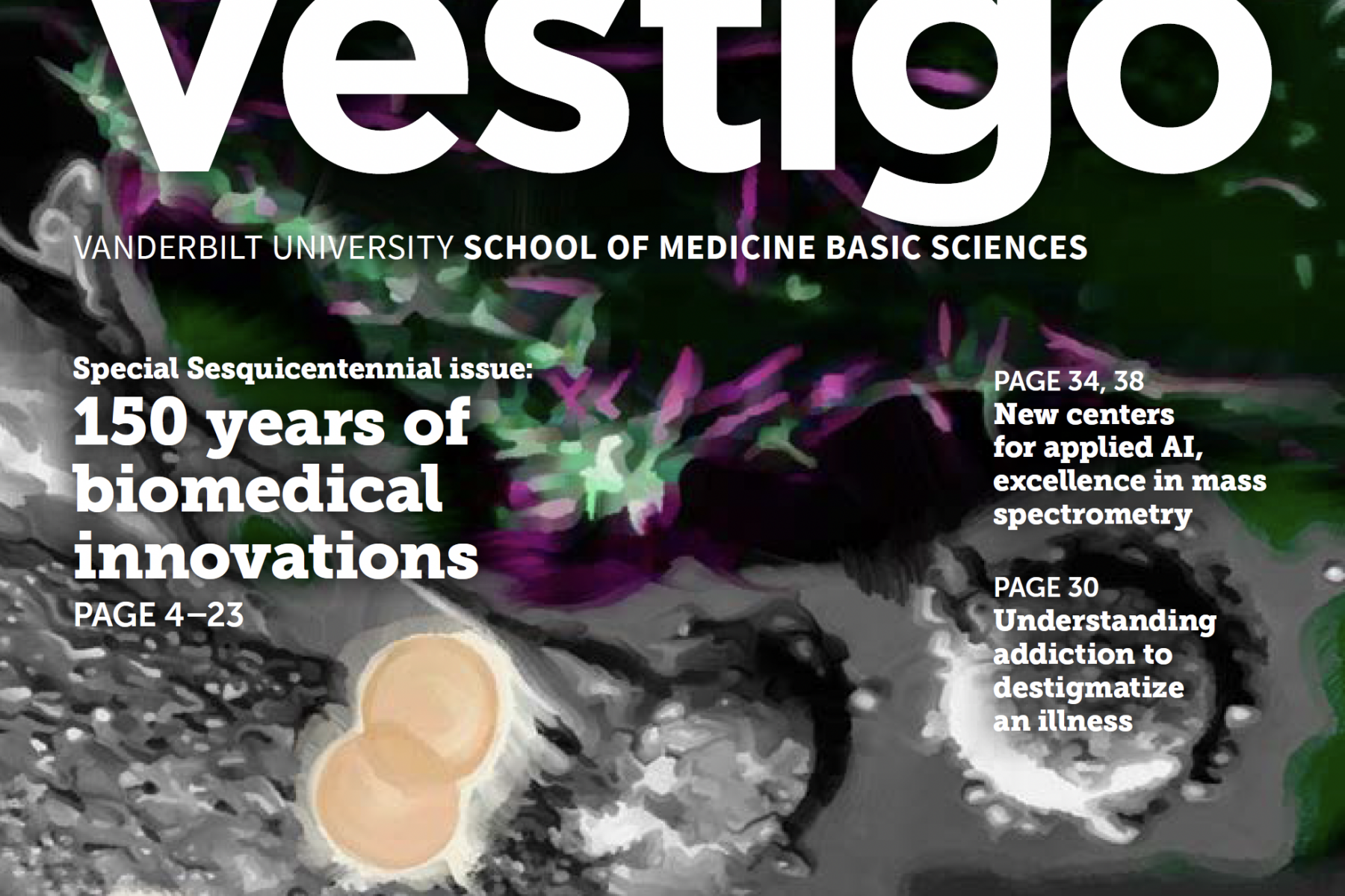
Thanks to an Microscopy Society of America Strategic Initiative grant, the Cell Imaging Shared Resource was able to purchase a new Zeiss Crossbeam 550 microscope, which will allow researchers across the Basic Sciences and the School of Medicine to do Focused-Ion-Beam Scanning Electron Microscopy. This image, taken by Evan Krystofiak, is the first-ever image taken on the new microscope and shows a basal lateral surface of enterocytes from the small intestine.
Featured in the May 2023 issue of Basically Speaking.
Zachary Sanchez – Dylan Burnette lab
This month’s banner image was taken by Zachary Sanchez, a Ph.D. student in the lab of Dylan Burnette (Cell and Developmental Biology. The image shows a human induced pluripotent stem cell-derived cardiomyocyte that is stained for alpha-actinin 2, a sarcomeric protein that localizes to z-lines and z-bodies. It is color coded to indicate depth (changes in the height of the cell). Sanchez took this proof-of-concept image while exploring the use of a new technique for his lab called expansion microscopy, which involves taking fixed cells or tissues and expanding their size to about four times their normal size. It is a method designed to get around the need for less accessible super-resolution techniques such as STORM.
Featured in the April 2023 issue of Basically Speaking.
Kendra Oliver – Pharmacology
The School of Medicine Basic Sciences will soon be launching its new named lecture series: Apex. The Apex series will feature researchers from around the world who are changing the topology of knowledge within their fields. This image is but one visual element that will accompany the Apex series, and it was selected and designed by Kendra H. Oliver (Pharmacology). This issue’s banner shows a variation on the theme of this image.
Featured in the March 2023 issue of Basically Speaking.
Ritika Raghavan – Danny Winder lab
This image was taken by Ritika Raghavan, a Ph.D. student in the lab of Danny Winder. Her research involves the study of alcohol and its effects on stress circuitry, which includes the paraventricular nucleus of the hypothalamus—pictured here. Cell nuclei are labeled in blue, neurons entering the PVH are labeled in red.
Featured in the February 2023 issue of Basically Speaking.
Anna Cassidy – Maria Žanić lab
Anna Cassidy, a Ph.D. student in the lab of Maria Žanić (Cell and Developmental Biology), generated a video while looking at microtubule networks in mitotic cells. This image was taken from the video and shows alpha-tubulin in green.
Featured in the January 2023 issue of Basically Speaking.
Elizabeth Orput – Rick Sando lab
The lab of Rick Sando (Pharmacology) focuses on understanding how synaptic connectivity between different neuronal populations self-assembles into functional neural circuits during development.
This image, taken by Sando lab research assistant Elizabeth Orput, shows mouse brain sections labeled for two mostly non-overlapping populations of inhibitory GABAergic neurons (red/green). The mutually exclusive pattern of these cells evokes the holiday spirit.
Happy holidays!
Featured in the December 2022 issue of Basically Speaking.
Michelle Bedenbaugh – Richard Simerly lab
This image was taken by Michelle Bedenbaugh, a postdoc in the lab of Richard Simerly. It shows melanocortin-3 receptor neurons (aqua) and Fos (red) in the brain. MC3R neurons that colocalize with Fos (a class of transcription factor) in this image are activated when animals are stressed.
Featured in the November 2022 issue of Basically Speaking.
Skylar Cuevas – undergraduate
Happy birthday, Oswald Avery! Avery, born on October 21, 1877, was the scientist who discovered that genes and chromosomes are made out of DNA. Avery died in 1955 and was buried in the Mount Olivet Cemetery in Nashville, where Dean Larry Marnett often takes his lab to commemorate him. This medallion was created by undergraduate Skylar Cuevas to distinguish Avery’s otherwise innocuous-looking gravestone. The medallion was placed during a Basic Sciences-hosted celebration of Avery’s 144th birthday in 2021.
Featured in the October 2022 issue of Basically Speaking.
Jessica Biddinger – Richard Simerly lab
This image is a still from a video of the “cleared” small intestine of a mouse with certain receptors marked with fluorescent proteins as observed through a light-sheet microscope (clearing makes tissue more transparent to visible light). The video was produced by Jessica Biddinger of the Richard Simerly lab as part of a project that’s looking at how the early postnatal environment can influence the development of neural circuits that are important for controlling metabolism. The Simerly lab is interested in how viscerosensory information from the gastrointestinal tract is conveyed to the brain.
Featured in the September 2022 issue of Basically Speaking.
Kendra Oliver
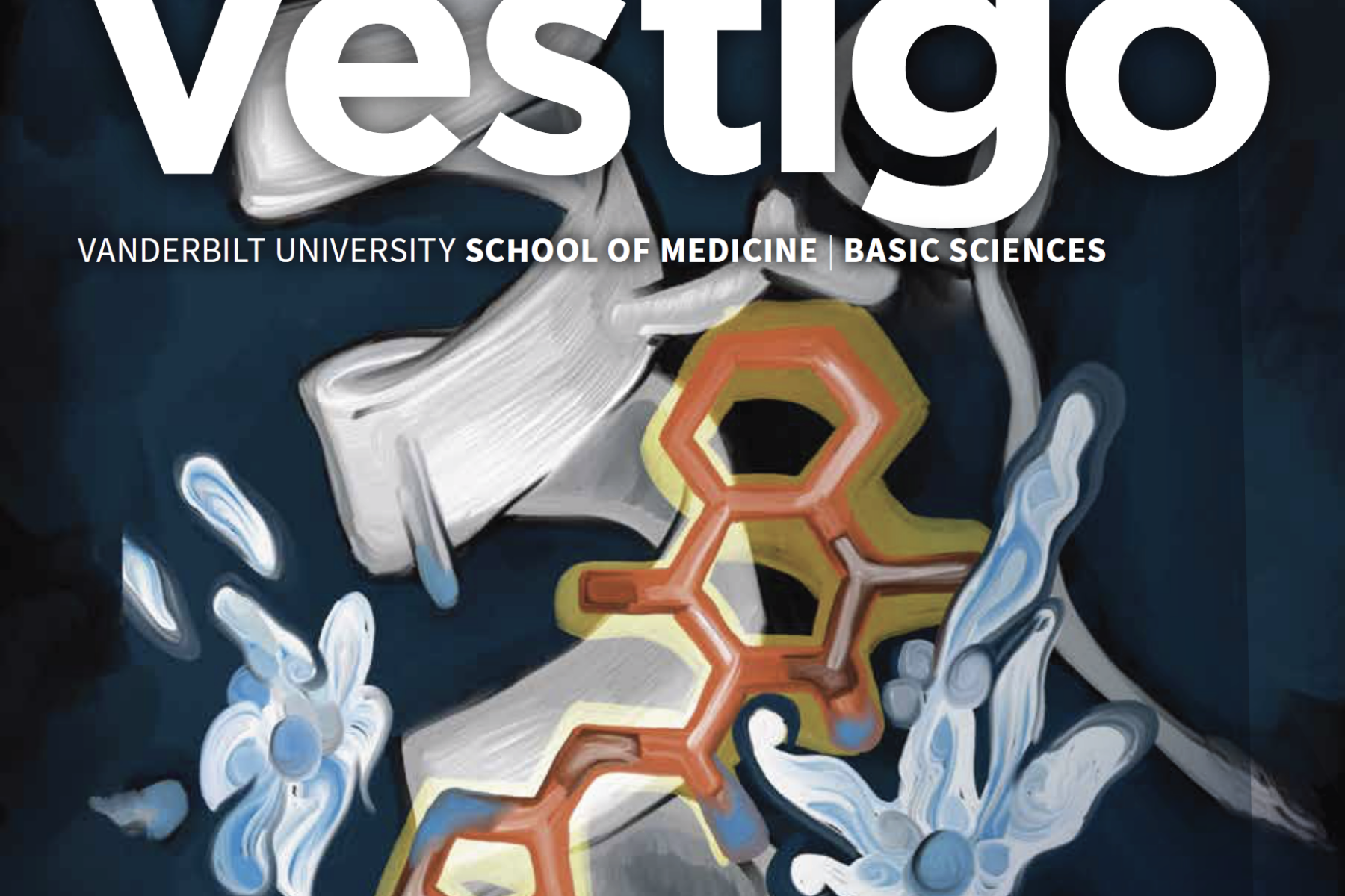
In celebration of Larry Marnett’s tenure as dean, this cover is dedicated to his work with COX-2, a human enzyme that is targeted by non-steroidal anti-inflammatory drugs. This image is based on the structure of the NSAID isoxicam when bound to COX-2, a structure that Marnett’s lab determined nearly eight years ago (2014). Water molecules mediate a crucial aspect of this interaction at amino acid residues R120 and Y385. Oxicams (including isoxicam) are the only class of NSAIDs for which water-mediated binding to COX-2 is observed. The structure, including the water molecules, is essential for explaining the structure-activity of this series of drugs. The illustration was made by Kendra Oliver (Pharmacology) and is the cover art for Issue 4 of Vestigo.
Featured in the August 2022 issue of Basically Speaking.
Julissa Burgos – Matt Tyska lab
Pathogenic Escherichia coli recruit the host cell protein actin to form structures that help the bacteria move around the cell surface. This image of HeLa cells was taken by Julissa Burgos, a graduate student in the lab of Matthew Tyska (Cell and Developmental Biology), as part of a study aiming to understand how host proteins play a role in pathogenic E. coli infection. DNA is shown in blue (the large blue spots are the host cell nuclei and the small blue spots are the bacteria) and F-actin is shown in purple.
Featured in the July 2022 issue of Basically Speaking.
Colleen Niswender – Warren Center for Neuroscience Drug Discovery
The artwork for this banner was created by Associate Professor of Pharmacology Colleen Niswender. This is what Niswender had to say about her painting titled “Neurons I Have Known and Loved”: “I enjoy creating abstract paintings that incorporate the feeling of movement. When this painting was complete, I realized it seemed to be a subconscious coupling of my scientific life as a neuropharmacologist with the artwork I’m currently exploring using the application of acrylic paint with palette knives. I hope you enjoy it!”
Featured in the June 2022 issue of Basically Speaking.
Matthew Roeske – Stephan Heckers lab
Matthew Roeske, an MSTP student in the Neuroscience graduate program, generated a surface reconstruction of the left human hippocampus from hundreds of structural MRI scans of patients with schizophrenia and healthy individuals. The surface is composed of 41,000 individual points, and each point has a specific coloration that indicates the magnitude of hippocampal shape deformation in schizophrenia patients at that precise location. Roeske works in the lab of Stephan Heckers (Psychiatry and Behavioral Sciences).
Featured in the May 2022 issue of Basically Speaking.
Vanderbilt University
This issue’s banner features a Vanderbilt University-branded mosaic with Giving Day donor-submitted photos. The mosaic will live on campus for years to come! Thank you to the more than 6,000 members of the VU community who supported scholarships, discovery, and belonging at Vanderbilt this Giving Day. Special shoutout to the 177 people who donated to Basic Sciences, helping to raise over $43,000 for our scientists! The money you donated, coupled with Dean Marnett’s and Dr. Mark A. Magnuson and Mrs. Lucile Magnuson’s matches, will help fund trainee programming, basic research grants, a new Brighter Ventures Student Award, the Stanley Cohen Innovation Fund, and more.
Featured in the April 2022 issue of Basically Speaking.
Aidan Fenix – Dylan Burnette lab
This image, by Aidan Fenix, a Ph.D. graduate from the lab of Dylan Burnette (CDB), is an electron micrograph taken as part of a project in collaboration with David Bader (Medicine, emeritus) that investigated a mutation in the CENP-F protein that resulted in dilated cardiomyopathy. This image shows the acto-myosin contractile machinery of WT mouse cardiomyocytes that has been pseudocolored to highlight the myosin (light magenta), actin filaments (orange), and mitochondria (dark purple blobs).
Featured in the March 2022 issue of Basically Speaking.
Alaina Willet – Kathy Gould lab
This image, by Alaina Willet, a postdoc in the lab of Kathy Gould (CDB), depicts live-cell imaging of fission yeast cells with labeled endocytic actin patches in cyan and the cytokinetic contractile ring and spindle pole bodies in magenta. These cells were blocked in the G2 stage of the cell cycle via a temperature-sensitive mutation, and were then released to proceed through mitosis. This is part of a project studying the function of myosin-1 at actin patches and the contractile ring in fission yeast.
Featured in the February 2022 issue of Basically Speaking.
Edgar Garza and Zer Vue – Antentor Hinton Jr. lab
This image is a 3D electron micrograph of mitochondria in a cell (each mitochondrion is in a different color), taken from a video reconstruction made by Edgar Garza and Zer Vue, members of the lab of Antentor Hinton Jr. (MPB), was generated as part of a recent preprint. Written in collaboration with first author Gabriella Robertson and the lab of Vivian Gama (CDB), the preprint focuses on how dynamin-related protein 1-mediated mitochondrial fission is essential to maintaining cristae morphology and bioenergetics.
Featured in the January 2022 issue of Basically Speaking.
Kathy DelGiorno – Kathy DelGiorno lab
This image of a metaplastic duct in the pancreas shows lineage-traced acinar cells in green and tuft cells in red. The co-immunofluorescence image was taken by the lab of Kathy DelGiorno as part of a project that’s looking at acinar-to-ductal metaplasia, as ADM occurs in the pancreas in response to tissue injury and is a potential precursor for adenocarcinoma. The cells were, of course, decorated for the holidays!
Featured in the December 2021 issue of Basically Speaking.
Kendra Oliver
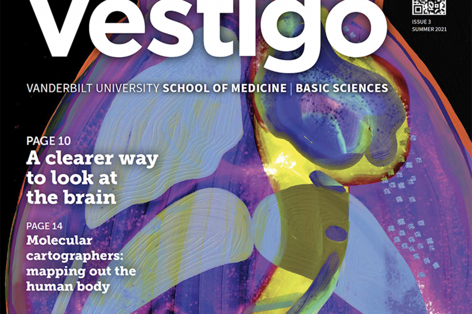
An artistic interpretation of a two-dimensional brain representation generated by José Maldonado through whole-brain imaging carried out at the Vanderbilt Neurovisualization Lab. The illustration was made by Kendra Oliver (Pharmacology) and is the cover art for Issue 3 of Vestigo.
Featured in the November 2021 issue of Basically Speaking.
Göker Arpağ – Marija Žanić lab
This image, generated by postdoc Goker Arpag of Marija Žanić’s lab, is a temporally color-coded representation of the movement of microtubule polymers on a surface coated with kinesin-1 motors. For this “gliding assay,” the kinesins — motor proteins that walk along microtubules — are attached to the surface of a coverslip via their cargo-binding domain, and microtubules are added to the flow-cell on top of the kinesins. As the immobilized kinesins bind and walk along microtubules, microtubules ‘glide’ over the coverslip. Addition of proteins that serve as barriers to kinesin-based transport disrupts microtubule gliding, resulting in the microtubules circling around a pivot point. In vitro reconstitution assays like this one allow researchers to dissect the molecular mechanisms underlying kinesin-based transport in cells.
Featured in the September 2021 issue of Basically Speaking.
Laura Geben – Rebecca Ihrie lab, and Serena Sweet – Richard Simerly lab
Immunolabeling-enabled three-dimensional imaging of solvent-cleared organs (iDISCO+) is a technique used to clear and immunostain organs, tissues, and even whole embryos. To obtain this image, Laura Geben (Pharmacology, Rebecca Ihrie lab) and Serena Sweet (MPB, Richard Simerly lab) used iDISCO+ to clear an embryonic mouse on day 13.5. The embryo was stained with an antibody against tropomyosin receptor kinase A (TrkA), a marker of the developing nervous system. After clearing, the embryo was then imaged on a light sheet microscope at the newly established Neurovisualization Lab.
Featured in the August 2021 issue of Basically Speaking.
Maya Igarashi – Kathy Gould lab
This image of mutant fission yeast cells was taken by Maya Igarashi, a research assistant in the lab of Kathy Gould (CDB). These mutant cells abnormally accumulate cell wall material at the septum (middle) and tips. Igarashi snapped this image for a study exploring cell wall construction in fission yeast, and stained the cells with the cell wall stains calcofluor (cyan) and FITC-lectin (magenta).
Featured in the June 2021 issue of Basically Speaking.
Colbie Chinowski – Matt Tyska lab
This image, taken by Colbie Chinowski (CDB), a graduate student in the lab of Matt Tyska, shows a network of non-muscle myosin 2C, a protein expressed only in the inner ear, the intestines, and the pituitary gland, stretching across the apical surface of enterocytes (intestinal absorptive cells). Chinowski’s research focus has been on understanding the function and organismal localization of NM2C, which remains uncharacterized despite its very strong enrichment in the intestine.
View larger image.
Featured in the April 2021 issue of Basically Speaking.
Robert Lavieri – VICTR
To cap the year, we’re featuring art by VICTR Senior Project Manager Robert Lavieri. He uses a program called UCSF Chimera to render images from coordinates in the Protein Data Bank. He then takes that “raw” image and modifies it in Photoshop to create art like this piece. Here, Lavieri worked with 5DE5, the complex between the RGG motif of the human Fragile X mental retardation protein and G-quadruplex RNA. FMRP is a regulatory RNA binding protein that plays a central role in the development of several human disorders including Fragile X Syndrome and autism. In this image, you can see both the protein — a yellow ribbon — and a stretch of bound RNA — in rainbow colors. The red spheres are potassium cations.
View larger image.
Featured in the December 2020 issue of Basically Speaking.
Fei Yang – Seth Bordenstein lab
This art piece was created by an Artlab Artist-in-Residence, Fei Yang, who worked in the lab of Seth Bordenstein in 2019. The image is meant to represent the work done in the Bordenstein lab, which focuses on animal-microbe associations. The teal portion represents Wolbachia-infected fruit fly testes, and the viral particles represent viral infections of the Wolbachia themselves. Wolbachia is a bacterial genus that infects many arthropod species and is perhaps the most common parasitic microbe on the planet. The nested symbiosis is represented by multiple layers of the cell separated by semi-circular membranes.
View larger image.
Featured in the October 2020 issue of Basically Speaking.
Amy Stark – John Penn lab
Graduate student Amy Stark (Pharmacology) has been working on optimizing the growth conditions of retinal pigment epithelial cells. Although she expected to find an even monolayer of cells when she looked under the microscope, she instead found isolated cells in the shape of—of all things—Gonzo the Muppet! Although the immunocytochemistry she ran to validate the RPE cells’ identity did not show the expected tight junctions that would have indicated a monolayer, she got a kick out of seeing her cells. And just like Stark, we think it’s important to find the joy in the day-to-day of science, even when things don’t go right. View larger image.
Featured in the September 2020 issue of Basically Speaking.
Kendra Oliver
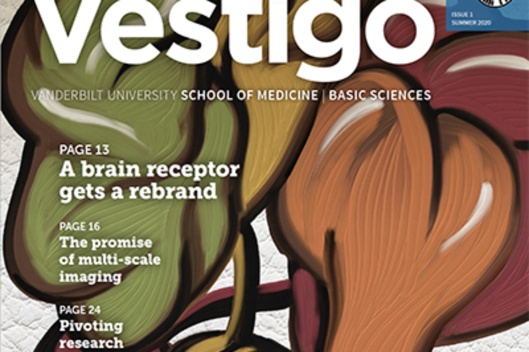
The cover of Vestigo and the banner for this month’s newsletter was drawn by Kendra Oliver, an assistant professor of pharmacology. She depicted an AMPA receptor, a glutamine receptor in the brain and the focus of study of Teru Nakagawa. A faculty member in Molecular Physiology and Biophysics, Nakagawa’s work is highlighted in Vestigo’s first issue. Oliver’s science background and artistic talent allows her to represent scientific concepts in a visually stunning manner. View larger image.
Featured in the July 2020 issue of Basically Speaking.
Greg Salimando – Danny Winder lab
The image shown here was taken by Greg Salimando, a recent graduate from the lab of Danny Winder. It shows how mRNA transcripts for the N-methyl-d-aspartate receptor (NMDAR) subunit GluN2D prominently co-localize with transcripts for the neuropeptide corticotropin releasing factor (CRF) in the bed nucleus of the stria terminalis (BNST). This 20X image of the mouse BNST shows individual neural cells counterstained with the DAPI nuclei stain, in blue, and labeled for transcripts of both GluN2D (red) and CRF (green). Greg’s research during his time in Dr. Winder’s lab concerned whether GluN2D-NMDA receptor activity in the BNST could regulate emotional behavior. See his recent paper (NMDA receptors are an appealing therapeutic target, above) for more information. View larger image.
Featured in the June 2020 issue of Basically Speaking.
Kit-Yi Yam and Jose Maldonado – Richard Simerly lab
The image shown here is a forebrain cross section derived from a 3D reconstruction of light-sheet fluorescence images of an optically cleared mouse brain. Using a technique called iDISCO+, postdoc Kit-Yi Yam and Research Instructor Jose Maldonado (Richard Simerly lab, MPB) optically cleared a mouse brain and immunohistochemically labeled it for an axonal marker (green) and a neuronal marker (red). They then imaged the entire brain to reveal the distribution of axonal projections from a subpopulation of hypothalamic neurons involved in feeding. View larger image.
Featured in the May 2020 issue of Basically Speaking.
Chuck Sanders – Chuck Sanders lab
The Sanders lab is devoted to characterizing the structures, folding and misfolding, and molecular mechanisms of membrane proteins using a variety of biochemical and biophysical methods. This artwork was created by biochemistry faculty Chuck Sanders, and features the structures of a variety of membrane proteins relevant to his lab’s work. The background is a photo Sanders took of watercress growing in the crystal-clear Ninfa river in Italy, where the Romans believed river nymphs lived. View larger image.
Featured in the March 2020 issue of Basically Speaking.
Amy Kendall – Lauren Jackson lab
Retromer is a protein complex found in endosomes that can load and ferry different kinds of cargoes to different cellular locations. This cryo-EM image of retromer shows it in a chain assembly formation. Image taken by Amy Kendall, lab manager of the Lauren Jackson lab. View larger image.
Featured in the February 2020 issue of Basically Speaking.
Noel Maxwell – Walter Chazin lab
2D NMR plot of wild-type calmodulin (black) and a mutant (E104K, red). Calmodulin is a calcium-binding messenger protein that is critical for Ca2+ signaling, and the E104K mutation causes a dysfunction in calcium binding. Although most mutations are embryonic lethal, this particular mutant was identified in a little girl who had recurrent cardiac arrests caused by arrhythmia. This NMR plot was generated by Noel Maxwell, a former research assistant in the lab of Walter Chazin (Biochemistry). View larger image.
Featured in the January 2020 issue of Basically Speaking.
Eric Figueroa – Jerod Denton lab
Eric Figueroa, from the lab of Jerod Denton, studies VRAC, an ion channel that conducts Cl– and organic osmolytes out of cells to draw water out. In the lab, he exposes cells to a hypotonic solution to cause swelling, using a pipet to create a gradient. When swelling, the cell membrane detaches from the cytoskeleton, creating blebs (black arrow). View larger image.
Featured in the December 2019 issue of Basically Speaking.
Kristin Peterson – Julie Rhoades lab
Some cancers metastasize specifically to the bone, and Kristin Petersen, a graduate student in the lab of Julie Rhoades, part of Vanderbilt’s Center for Bone Biology, studies one such cancer: oral squamous cell carcinoma (OSCC). OSCC affects the cells that line the lips and the inside of the mouth and can invade the bones of the mandible. The image shows some cetuximab-resistant OSCC cells, which are more elongated and are more prone to migrating than non-cancer cells. The cells are stained in red (lipophilic dye), and their nuclei in blue (DAPI). View larger image.
Featured in the November 2019 issue of Basically Speaking.
Chris Hofmann – Ron Emeson lab
Chris Hofmann, a grad student in the lab of Ron Emeson, studies a G protein-coupled receptor (GPCR) called mGlu4. This image shows a calcium-sensing assay that’s used to indirectly measure the activity of mGlu4. Each spot on this assay turns darker or lighter depending on the level of activity of the GPCR in each spot. When measured over time, Hofmann can see the intensity of the light increase and decrease. He is currently exploring how point mutations affect mGlu4’s function. View larger image.
Featured in the October 2019 issue of Basically Speaking.
Romell Gletten – Kevin Schey lab

Romell Gletten is a graduate student in the Kevin Schey lab. He studies the mechanisms that mediate the cellular trafficking of critical proteins such as the aquaporins in lens fiber cells. This includes the investigation of these mechanisms under both normal physiology and cataractogenesis, in addition to relevant modulatory post-translational modifications. Romell primarily employs biochemical techniques, including mass spectrometry-based proteomics, and molecular biological methods to address this research focus in both humans and animal models. View larger image.
Featured in the September 2019 issue of Basically Speaking.
Alexandria Oviatt – Neil Osheroff lab
Alexandria Oviatt, a graduate student in the lab of Neil Osheroff, works with bacterial topoisomerases, enzymes that regulate supercoiling and that can remove knots and tangles from DNA. This image is a 2D gel that she generated after testing the ability of a Staphylococcus aureusgyrase (a type II topoisomerase that can introduce negative supercoils) to relax DNA. DNA can be electrophoresed on agarose gels to discern its supercoiling state, but positively and negatively supercoiled DNA run at similar speeds. To get around this problem, Oviatt runs her gels in two directions, which helps separate the different DNA species. This 2D gel shows a time course in which the gyrase relaxed positively supercoiled DNA (top left) before introducing negative supercoils (bottom right). Image courtesy of Alexandria Oviatt and Elizabeth Gibson. View larger image.
Featured in the August 2019 issue of Basically Speaking.
Caroline Cencer – Matt Tyska lab
There is more to this artwork than meets the eye: this kaleidoscopic image is actually a single immunofluorescence slide that has been rotated three times about a corner. The artist and researcher, graduate student Caroline Cencer (lab of Matt Tyska), uses enterocytes to study the maturation of the intestinal brush border. In this image, the microvilli (magenta) are coming out of the screen toward the viewer. View larger image.
Featured in the July 2019 issue of Basically Speaking.
Caroline Roe and Justine Sinnaeve – Rebecca Ihrie and Jonathan Irish labs

This image shows a glioblastoma tumor that was frozen, sectioned, and stained with metal-tagged antibodies; 7 (out of more than 30) markers are shown in this particular visualization. This image is exemplary of a pilot project between the labs of Rebecca Ihrie and Jonathan Irish to bring imaging mass cytometry (IMC) to Vanderbilt. IMC allows researchers to identify 30-35 different markers to get a nuanced understanding of the types of cells that are present in a single sample, like with mass cytometry, except that IMC allows them to discern structural and spatial relationships between cell subsets. Caroline Roe is a program manager in the Irish lab and Justine Sinnaeve is a graduate student in the Ihrie lab. View larger image.
Featured in the June 2019 issue of Basically Speaking.
Kensei Taguchi – Craig Brooks lab
Autophagosomes in kidney tissue using an RFP-GFP-LC3 reporter mouse. Shown in super-resolution structured illumination microscopy (SIM). Tissue sections were stained for GFP (green), RFP (red), KIM-1 (magenta), and DAPI (blue). View larger image.
Featured in the May 2019 issue of Basically Speaking.
Caitlin Sprowls – Seth Bordenstein lab
Histological sections of California sea lion whiskers, trichrome stained and then color manipulated in photoshop. Image courtesy of ArtLab, taken by Caitlin Sprowls of the Seth Bordenstein lab. View larger image.
Featured in the April 2019 issue of Basically Speaking.
Amrita Pathak – Bruce Carter lab
Primary sympathetic neurons cultured in microfluidic devices. Cells are stained with neuronal marker TUJ1 (green) and nuclei are labeled with DAPI (blue). Submitted by Amrita Pathak, Research Instructor in the lab of Bruce Carter. (Image courtesy of ArtLab.) View larger image.
Featured in the March 2019 issue of Basically Speaking.
Nilay Taneja – Dylan Burnette lab
Embryonic Stem Cell Colony
Shown is a colony of human embryonic stem cells showing the actin cytoskeleton (magenta), myosin motors (cyan) and DNA (yellow). Embryonic stem cells, that give rise to all tissues in the body, hold great potential in designing cell-based therapies for multiple diseases. Studying the cytoskeleton of these cells helps us understand how they respond to their mechanical environment and how that affects their ability to both retain their identity or differentiate into specific cell types. Technique used- Confocal microscopy, large image stitching.
Image taken by Nilay Taneja, a graduate student in the Dylan Burnette lab. View larger image.
Featured in the February 2019 issue of Basically Speaking.