By Colbie Chinowsky

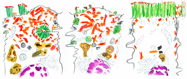
A collaboration between investigators at Vanderbilt University and the University of Arizona has revealed new insights about a rare, genetic neonatal ailment, microvillus inclusion disease (MVID), which affects the enterocytes of the small intestine. This work, published in the Journal of Cell Biology and led by James Goldenring (Surgery) in collaboration with Matt Tyska (Cell and Developmental Biology), uncovered part of the mechanism that leads to MVID, and opened up new questions about what makes it deadly.
Enterocytes are the only nutrient-absorbing cells in the body and have evolved to maximize their absorption abilities. The brush border, composed of thousands of protrusions (microvilli) that extend from cells outward into the intestinal tube, allows for a 9-16-fold increase in nutrient absorption thanks to an increase in surface area. Maintenance of microvilli is critical for homeostasis, and many diseases that affect microvilli result in intestinal distress and malnutrition.
Myosin Vb, a motor protein that helps determine cell polarity or asymmetry, has been previously implicated in MVID. A lack of myosin Vb function changes a cell’s polarity and results in the formation of small clumps of microvilli forming inside the cell, where they mix with digestive proteins to form structures called microvillus (singular of microvilli) inclusions. Such inclusions are the hallmark of MVID, which results in life-threatening malnutrition due to the lack of proper nutrient absorption machinery.
To date, the origins of the inclusions in MVID remain unknown, but this research clarifies which of the cellular processes get hijacked to allow for inclusion formation. The researchers found that knocking out the gene for myosin Vb in mice results in the formation of inclusions. Studying these mice allowed them to determine how these inclusions are formed, and what proteins are necessary.
“This paper answers a contested question in the field: what are these inclusions, and are they formed via endocytosis or exocytosis?” said Amy Engevik, a postdoctoral fellow in the Goldenring lab and the first author on this paper.
Endocytosis is a process in which a cell engulfs outside matter, and exocytosis one where the cell expels matter into its environment.
Using small intestinal tissue from these mice, Goldenring and colleagues grew enteroiods – spherical structures grown from intestinal stem cells that recapitulate the architecture of the intestine – and monitored the brush border using live fluorescence microscopy. Using this technique, they were able to observe inclusions form; however, they wanted to know more about how they formed, believing that endocytosis might be involved. After adding a large fluorescent dextran, a sugar molecule, to the enteroiods’ media, they saw it inside the inclusions, suggesting that these enterocytes had absorbed the sugar and the microvilli along with other outside matter through bulk endocytosis (regular endocytosis would not have been capable of absorbing such large cargo).
Bulk endocytosis is known to occur in hyper-stimulated neurons, a process termed activity-dependent bulk endocytosis (ADBE). The authors found that bulk endocytosis in enterocytes required similar proteins to ADBE, including dynamin, a protein that helps cut endocytic vesicles or pouches once they bring in material from the outside of the cell. They speculate that bulk endocytosis in the small intestine may normally help to rapidly absorb material early in life. In the case of a myosin Vb knockout mouse, which mimics the Vb mutations seen in MVID patients, the process gets co-opted to produce inclusions.
Surprisingly, however, when they examined a double-knockout of myosin Vb and Pacsin2, a protein involved in endocytosis, the authors found that although the number of inclusions decreased, the mice were still very sick. This indicates that although the inclusions are a hallmark of MVID, they are not the reason for the deadly diarrhea and malnutrition these patients experience.
“The inclusions were a bit of red herring,” concluded Engevik.
To begin understanding how to treat this disease, more research is needed into how myosin Vb causes the symptoms of MVID, as the inclusions appear to merely be a side effect. This study represents one step toward helping patients affected by this rare disease.