
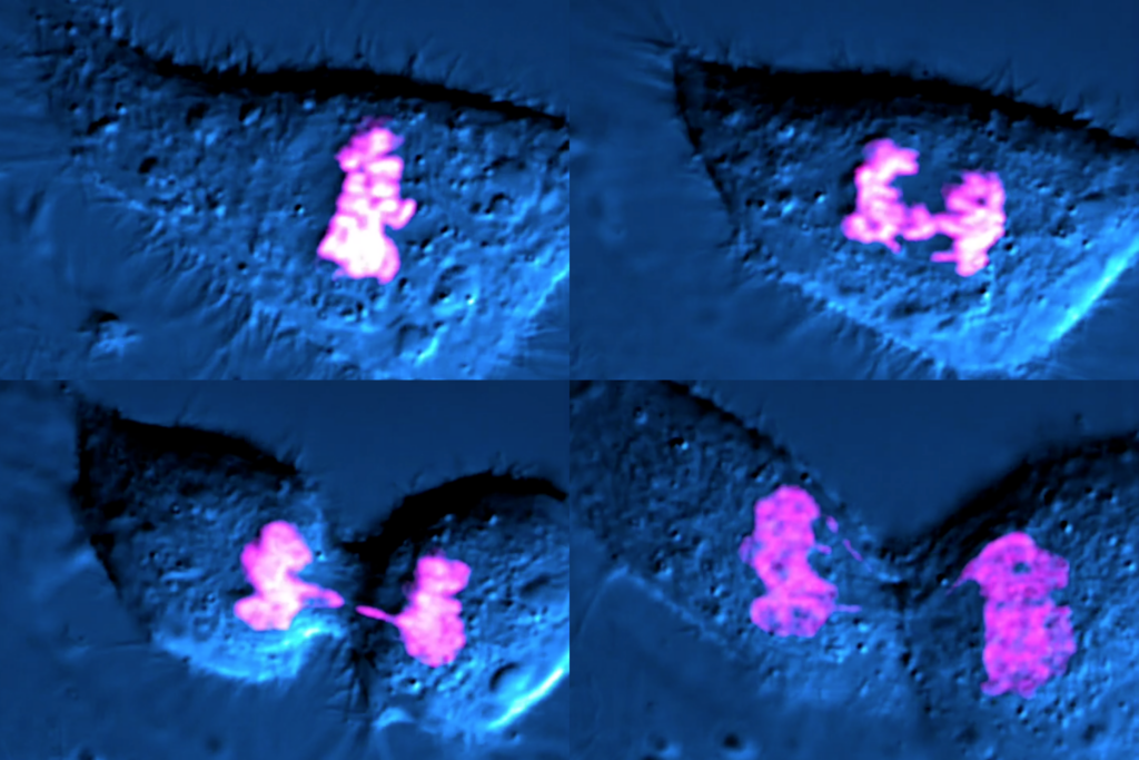
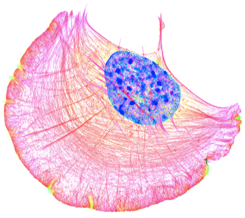
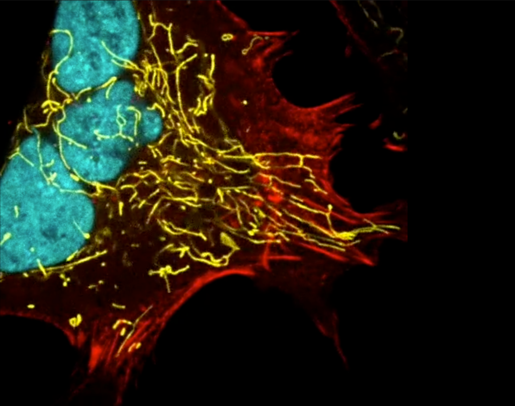
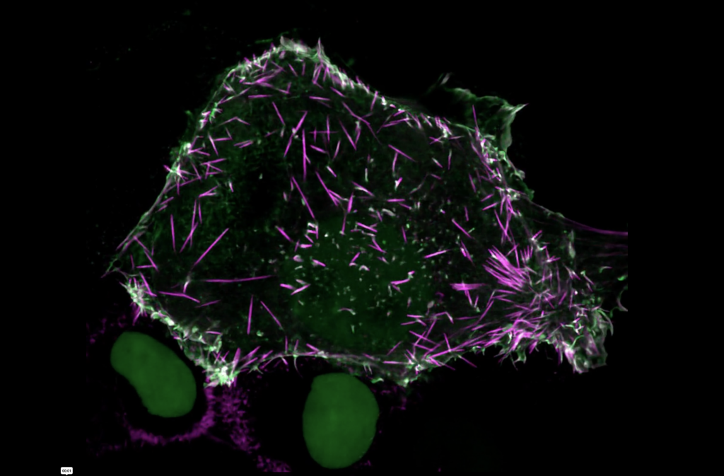
Dylan Burnette, associate professor of cell and developmental biology, and Olivia Perkins, a graduate student in the Department of Cell and Developmental Biology, were awarded prizes in the 2022 Nikon Small World and Nikon Small World in Motion competitions.
Founded in 1975, Small World recognizes the top microscope images and movies by annually awarding winning and honorable mention prizes. Aptly summarized by comedian Stephen Colbert as “a contest for tiny pictures of tiny things,” Small World has become legendary and serves as a visual testament to the advancement of microscopy for nearly 50 years.
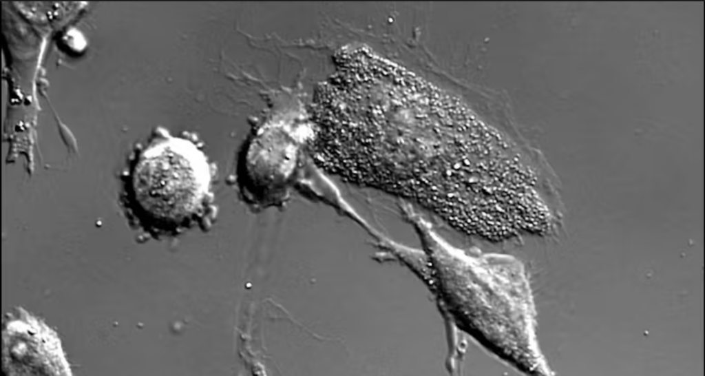
Burnette was awarded one honorable mention in the image category, in addition to fourth place and two honorable mentions in the movie category. Perkins was awarded an honorable mention in the movie category.
Despite the vast number of submissions to the contest from scientists worldwide each year, Vanderbilt is no stranger to Small World awards. Vanderbilt researchers have amassed a total of 17 recognized images and movies in the past several years, placing Vanderbilt in the top 15 of the most awarded institutions in the history of the competition.
Vanderbilt’s success can be attributed in part to the outstanding microscopy core facilities available on campus. Led by Scientific Director Matthew Tyska, who holds a Cornelius Vanderbilt Chair and is a professor of cell and developmental biology, and Managing Director Jenny Schafer, who is also a research associate professor of cell and developmental biology, the Cell Imaging Shared Resource offers a wide variety of microscopes—including super-resolution and electron microscopes—across campus.
In 2016, CISR opened its state-of-the-art Nikon Center of Excellence facility in partnership with Nikon Instruments. By housing microscopes within five suite locations, CISR has established an easily accessible imaging resource for students and staff. CISR personnel are always on hand to train researchers on the proper use of the microscopes as well as assist in data processing and analysis.
CISR keeps up with the newest microscopes and techniques. Most recently, CISR acquired a lattice light-sheet microscope and a focused ion beam scanning electron microscope. The light-sheet microscope enables fast, three-dimensional imaging of live cells without photo-toxicity—which happens when too much excitation light damages the cell—thanks to thin sheets of light passing through the sample one slice at a time. The addition of the FIB-SEM is especially exciting, as this new technology applies the high-resolution power of SEM to the three-dimensional space, allowing CISR users to create in-depth maps of entire cells or subcellular structures.
Burnette and Perkins made use of CISR microscopes—including the structured illumination microscope and the spinning disk confocal microscope—and training, which set them up for recognition by Small World. Many of CISR’s resources are funded by shared instrumentation grants or center grant supplements as noted in the numerous scientific publications that recognize the use of the CISR core each year. As CISR continues to acquire the latest and greatest microscopes, the future of Vanderbilt imaging is truly limitless.