Apex Lecture Series
There are major inflection points in biomedical discovery that create new fields, new ideas, and new opportunities to impact human health. To engage with global researchers contributing to these inflection points, the Vanderbilt School of Medicine Basic Sciences launched the Apex Lecture Series in 2023. This school-wide seminar series brings scientists who are influencing the trajectory of their fields to engage with our scientific community on campus.
Apex Lectures
-
2025 speakers
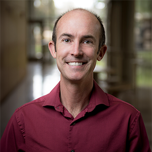 Ryan Hibbs, University of California, San Diego
Professor and Chair of Neurobiology, Professor of Pharmacology"Muscle Acetylcholine Receptors in Development and Disease"
Thursday, March 27, 4:00 p.m., 1220 MRB III
A reception will follow the lecture at 5:00 p.m. in the MRB III first-floor atrium.
Co-sponsored by the Department of Pharmacology and the Center for Applied AI in Protein DynamicsAbstract:
We are broadly interested in how ion channels enable fast signaling across membranes. This signaling underlies diverse physiological processes, from chemical transmission in the brain, to muscle contraction, to environmental sensation. In the seminar, I will discuss recent work from our lab on how connections between our nervous system and skeletal muscle develop, and how, in cases of autoimmune disease, these junctions are attacked. I will cover how we purified micrograms of muscle receptor protein from kilograms of bovine muscle (beef) to yield high-resolution structures of the acetylcholine receptor, then used electrophysiology to understand development of this synapse. In the second part of the seminar, I will show how we are using structural and functional methods to map epitopes for pathogenic patient antibodies and understand mechanisms underlying the autoimmune disease myasthenia gravis.
About the speaker:
Hibbs is the chair and a professor of neurobiology and a professor of pharmacology in the School of Biological Sciences at the University of California, San Diego. He studied chemistry and biochemistry at Whitman College, a small liberal arts college in Washington State, graduating in 2000. He did his Ph.D. research at UC San Diego with Palmer Taylor between 2001–2006 and his postdoctoral research with Eric Gouaux at the Vollum institute between 2007–2012. Hibbs started his independent lab in 2012 in the departments of neuroscience and biophysics at UT Southwestern Medical School, and was promoted to associate professor in 2019. In 2023 he moved his research lab to UC San Diego, but he maintains an adjunct appointment in neuroscience at UT Southwestern.
Awards and honors include Klingenstein and McKnight Scholar awards, the UC San Diego Outstanding Alumnus Award, the Alton Ochsner Award Relating Smoking and Disease, and the Norman Hackerman Award in Chemical Research.
The Hibbs lab investigates the fine details of fast communication in the nervous system, how this signaling goes awry in disease, and how drugs alter it. Ongoing projects span basic biophysics and structural biology, neuropharmacology, mechanisms underlying autoimmune diseases of the nervous system, and the evolution of neurotransmitter receptors. The laboratory is pursuing structural mechanisms of neurotransmitter receptor function and relationships to disease pathology, with a current focus on nicotinic acetylcholine receptors and GABAA receptors. We bring complementary methods to bear on the study of these receptors, including structural biology, pharmacology, and electrophysiology, and frequently collaborate with simulation experts, synthetic chemists, and clinicians. Long-term goals include understanding neurotransmitter receptor evolution, defining details in how they are targeted in autoimmune diseases, and enabling drug design relevant to addiction, mental illness, epilepsy, autoimmunity, and neurodegeneration.
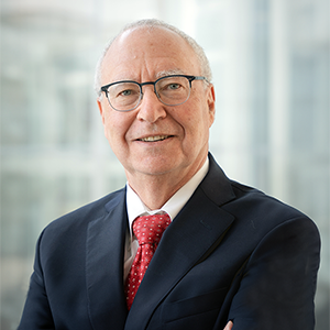 Mitch Lazar, University of Pennsylvania Perelman School of Medicine
Willard and Rhoda Ware Professor of Diabetes and Metabolic Diseases
Director, Institute for Diabetes, Obesity, and Metabolism"Nuclear Receptors and the Circadian Regulation of
Metabolism"Tuesday, Feb. 11, 3:00 p.m., 1220 MRB III
A reception will follow the lecture at 4:00 p.m. in the MRB III first-floor atrium.
Co-sponsored by the Department of Molecular Physiology and Biophysics.Abstract:
Circadian rhythms are an inherent feature of mammalian physiology, controlled by molecular clocks comprised of transcriptional and translational feedback loops that have evolved to anticipate the 24-hour light-dark cycle on Earth. Environmental challenges such as shiftwork or jetlag are detrimental to metabolic health, but how desynchronous signals are transmitted among tissues is not well understood. REV-ERB nuclear receptors are key components of the molecular clock and a major link between circadian rhythms and metabolism.
We have used conditional deletion strategies to address the role and hierarchies of the molecular clocks in different cells and tissues, focusing on the liver, which is central to systemic metabolism, as well as the body's master clock, located in the suprachiasmatic nucleus of the hypothalamus. Mice lacking REV-ERBs in hepatocytes exhibit rhythmic changes in lipid metabolism due to cell-autonomous loss of diurnal expression of a subset of liver genes, as well as non-cell-non-autonomous alterations of rhythmic gene expression in other liver cell types. Loss of SCN REV-ERBs leads to a marked shortening of behavioral rhythms and a desynchrony with the 24-hour light-dark cycle that predisposes to metabolic dysfunction, including fatty liver. Unexpectedly, hepatic REV-ERBs also provide feedback to the brain via the vagus nerve to control of food intake. Together the results help explain the metabolic disruption resulting from desynchrony between environmental conditions and endogenous clocks.
About the speaker:
Lazar is the Willard and Rhoda Ware Professor of Diabetes and Metabolic Diseases and the director of the Institute for Diabetes, Obesity and Metabolism at the University of Pennsylvania. He received an S.B. in chemistry from the Massachusetts Institute of Technology and an M.D. and Ph.D. from Stanford University. After training in internal medicine at Brigham and Women’s Hospital and endocrinology at Massachusetts General Hospital, Lazar joined the faculty of the University of Pennsylvania. He served for 24 years as chief of the Division of Endocrinology, Diabetes and Metabolism and is the founding director of UPenn's Institute for Diabetes, Obesity and Metabolism. Lazar's groundbreaking research has focused on the circadian rhythms and metabolism, and he has made fundamental contributions to the fields of endocrinology, diabetes, and chronobiology.
Lazar has served as a member of the Board of Scientific Councilors of the National Institute of Diabetes and Digestive and Kidney Diseases as well as many editorial and scientific advisory boards. He has been elected to the American Society for Clinical Investigation cand the Association of American Physicians and their respective councils, and served as president of the AAP Council in 2020–2021. He has received numerous awards from international societies and universities, including the Stanley J. Korsmeyer Award from the ASCI, the Transatlantic Medal from the Society for Endocrinology, the Roy Luft Award from the Karolinska Institute, the Keith Harrison Memorial Lectureship from the Endocrine Society of Australia, and the Fred Conrad Koch Lifetime Achievement Award from the Endocrine Society. He also received the 2025 George M. Kober Medal from the AAP for his pioneering and enduring, impactful contributions to improve health. Lazar is an elected member of the National Academy of Medicine, the American Academy of Arts and Sciences, and the National Academy of Science.
 David Bartel, Whitehead Institute, Massachusetts Institute of Technology
Professor of Biology; Core Member, Whitehead Institute; Investigator, Howard Hughes Medical Institute"Regulation of mRNA Translation and Decay"
Wednesday, Jan. 15, 2:00 p.m., 1220 MRB III
A reception will precede the lecture at 1:00 p.m. in the MRB III first-floor atrium.
Co-sponsored by the Department of BiochemistryAbstract:
We investigate the molecular pathways that regulate gene expression by affecting the stability or translation of mRNAs. One interest is microRNAs, which are small regulatory RNAs that typically direct the destruction of their target transcripts. We have recently been studying the conformational changes required for microRNA-directed slicing of mRNAs. Another interest is mRNA poly(A) tails, which stabilize mRNAs in some contexts and increase translation efficiency in others. We have recently found the conditions required for tail length to influence translational efficiency and have defined major determinants of tail-length modulation (both lengthening and shortening) in the cytoplasm.
About the speaker:
David P. Bartel is a molecular biologist best known for his work on microRNAs. He is a professor of biology at the Massachusetts Institute of Technology, a member of the Whitehead Institute, and an investigator of the Howard Hughes Medical Institute. The Bartel lab is interested in the molecular pathways that regulate eukaryotic gene expression by affecting the stability or translation of mRNAs. The Bartel lab studies microRNAs and other small RNAs that specify the destruction and/or translational repression of mRNAs. They also study mRNA untranslated regions and poly(A) tails, and how these regions recruit and mediate regulatory phenomena. Bartel has helped to define microRNAs, their regulatory targets, and their molecular and regulatory consequences in animals and plants. In addition, his lab has made key contributions to the understanding of other RNA-silencing pathways of animals, plants, and fungi, the functions of mRNA poly(A) tails and 3′-untranslated regions, and the catalytic potential of RNA.
Bartel is committed to the education and training of young scientists. At MIT, he teaches an undergraduate course in evolution with Robert Berwick and a graduate course in nucleic acids with Ankur Jain. In addition, 35 Ph.D. students and 33 postdoctoral fellows have completed training in the Bartel lab, most of whom are thriving either as postdocs or as independent investigators.
-
2024 speakers

Agnel Sfeir, Memorial Sloan Kettering Cancer Center
PaineWebber Chair in Cancer Genetics"Two Genomes, One Balance: Mitochondrial and Nuclear DNA Stability"
Wednesday, October 30, 2024, 2:00 p.m. CT, 202 Light Hall
A reception precedes the lecture at 1:00 p.m. in 618 Light Hall.
Co-sponsored by the Department of BiochemistryAbstract:
Eukaryotes cells contain two distinct genomes – nuclear and mitochondrial – which have distinct evolutionary origins and are housed in separate environments. The interplay and coevolution of these genomes are crucial for maintaining cellular homeostasis. Perturbations to either genome can disrupt their crosstalk and affect cellular responses. I am fascinated by how cells manage these challenges to maintain the delicate balance of preserving genome stability while allowing genetic innovation.
My research program revolves around two fundamental questions related to genome stability. Firstly, we investigate the evolutionary benefits of the microhomology-mediated end-joining (MMEJ) repair pathway that fixes DNA breaks in the nuclear genome. Despite being widely conserved, MMEJ's physiological role remains elusive. We aim to test the hypothesis that MMEJ balances genome integrity and evolution by promoting extrachromosomal DNA circularization. The circularization of invading DNA through MMEJ is a protective mechanism that shields the host genome. These circularized elements can resist nuclease degradation and persist in the nucleoplasm, providing a substrate for potential integration and stimulation of genome diversity.
In another line of inquiry, we will explore how the nucleus manages perturbations to the mitochondrial genome. Aberrations in mitochondrial DNA profoundly impact cellular function. Yet, standard methods of assessing the extra-nuclear genome and efforts to study it in isolation fail to address fundamental questions about its instability. To tackle this, we will develop innovative genetic and molecular tools to investigate the outcome of three toxic mtDNA aberrations and determine how they alter mito-nuclear crosstalk.
About Dr. Sfeir:
Molecular biologist Agnel Sfeir studies how DNA is broken and repaired, and how these processes are related to aging, cancer, and other phenomena. Her scientific contributions are notable for their breadth and depth, unified by the theme of genome stability. Trained as a telomere biologist, she has made seminal contributions to our understanding of telomere protection and maintenance. Since establishing her lab in 2012, she has been fascinated by the remarkable ability of cells to preserve genomic stability and curious about how these pathways fail in cancer cells. Her laboratory has significantly advanced research in two key areas: DNA double-strand break (DSB) repair of nuclear DNA and mitochondrial DNA (mtDNA) stability.
A recent interest of hers is in the biology of the DNA inside of mitochondria — the “power plants” of the cell. She calls this DNA “the forgotten yet fascinating genome.” Dr. Sfeir joined Sloan Kettering Institute’s Molecular Biology Program in March 2021, having previously been a faculty member at New York University’s Skirball Institute of Biomolecular Medicine.

Photo by Kate Joyce and Santa Fe Institute Venki Ramakrishnan, MRC Laboratory of Molecular Biology, Cambridge, England
Group Leader"Why We Die"
Friday, September 27, 2024, 3:00 p.m., 208 Light Hall
A reception will immediately follow the lecture in Langford Lobby.Abstract:
Ever since we humans learned of our mortality, that knowledge has driven much of human culture. However, for most of our existence as a species, there was little we could do about aging and death. In the last few decades, this has changed. We are beginning to understand the biological basis of aging and how it leads to death. At the same time, longer life expectancies and reductions in fertility rates mean that societies face an increasingly aging population, with an urgent need to improve health in old age. The fear of aging and death is also driving a quest for radical life extension among some gerontologists. My book, on which this event is based, is an attempt to take a hard look at progress and prospects in the field of aging and also point out the social issues involved.
About Dr. Ramakrishnan:
Venki Ramakrishnan received his bachelor’s degree in physics from Baroda University in India in 1971 and his Ph.D. in physics from Ohio University in 1976. He then studied biology for two years at the University of California, San Diego before beginning his postdoctoral work with Peter Moore at Yale University. After a long career in the USA at Brookhaven National Laboratory and the University of Utah, he moved to England in 1999, where he has been a group leader at the MRC Laboratory of Molecular Biology in Cambridge. He received the Nobel Prize for Chemistry in 2009 and was the president of the Royal Society from 2015-20.
In 2000, Ramakrishnan’s laboratory determined the atomic structure of the 30S ribosomal subunit and its complexes with ligands and antibiotics. This work led to insights into how the ribosome “reads” the genetic code, as well as antibiotic function. Ramakrishnan’s lab subsequently determined high-resolution structures of functional complexes of the entire ribosome at various stages along the translational pathway, which led to insights into its role in protein synthesis during decoding, peptidyl transfer, translocation and termination. For the last decade, his laboratory has been applying cryoelectron microscopy to study eukaryotic and mitochondrial translation, especially initiation of translation and translational regulation.
Ramakrishnan is the author of two popular books, Gene Machine:The Race to Decipher the Secrets of the Ribosome (2018), a very frank popular memoir about the race for the structure of the ribosome, and more recently, Why We Die: The New Science of Aging and the Quest for Immortality (2024).
View a recording of this lecture (VUNet ID required).
Karen Adelman, Harvard Medical School
 Edward S. Harkness Professor of Biological Chemistry and Molecular Pharmacology
"Transcriptional Control of Stress and Immune Responses"
Wednesday, April 10, 2024, 2:00 p.m. CT, 1220 MRB III
A reception will immediately follow the lecture in the MRBIII atrium.
Co-sponsored by the Department of BiochemistryAbstract:
The transition of promoter-proximally paused RNA polymerase II (Pol II) to productive elongation is a central, regulated step in metazoan gene expression. At many genes, paused Pol II is released efficiently into the gene body by the kinase P-TEFb. However, paused Pol II can also be targeted for early termination by the Integrator complex. Importantly, Integrator acts at many stress-responsive genes, to attenuate their activity in the absence of stress signaling. Here, I will discuss the mechanistic and physiological consequences of disruptions in Integrator activity, which are implicated in inflammatory disorders, cancer and neurological disease.
About Dr. Adelman:
Dr. Karen Adelman is the Edward S. Harkness Professor of Biological Chemistry and Molecular Pharmacology at Harvard Medical School. She is also a Member of the Gene Regulation Observatory at the Broad Institute, and the Ludwig Cancer Center at Harvard Medical School. The Adelman lab pioneered genomic studies of RNA polymerase II (RNAPII) transcription, revealing that pausing of RNAPII in early elongation is a central regulatory step in metazoan gene expression. Ongoing work probes the interplay between transcription, RNA processing and epigenetic machineries to elucidate the determinants of mature mRNA formation in health and disease. Dr. Adelman received the NIEHS, NIH Early Career Award in 2006 and in 2010 earned the NIH Directors Award for Scientific Achievement. She was elected to American Academy of Arts and Sciences in 2023.
 Robert Tampé, Goethe University Frankfurt
Professor of Biochemistry at the Biocenter, Director of the Institute of Biochemistry"From Antigens to Defense: Cellular Machineries in Quality Control and Adaptive Immunity"
Thursday, March 7, 2024, 12:00 p.m. CT, 208 Light Hall
Co-sponsored by the Department of Molecular Physiology and BiophysicsAbstract:
To elicit an effective immune response against pathogens and cancerous cells, MHC I molecules undergo an eventful journey from their biogenesis in the endoplasmic reticulum to their final decoding by cytotoxic T cells on target cell surfaces. Cellular machines, consisting of transporters, chaperones, and receptor clients, facilitate MHC I biogenesis, assembly, quality control, and final recognition. However, the mechanistic integration of the corresponding processes remains poorly understood. I will discuss the multichaperone-client interaction network of the MHC I peptide loading complex assembled on the transporter associated with antigen processing. By integrative approaches, we reveal the mechanistic underpinnings of antigen transport, epitope proofreading, quality control, and final release of MHC I complexes. In addition, the structural and mechanistic insights into the T cell receptor complex bound to a tumor-specific human class I pMHC will be discussed, including the functional impact of connecting peptides and sterol lipid for complex assembly.
 Xuewu Zhang, UT Southwestern Medical Center
Professor, Department of Pharmacology, Department of Biophysics"Structures of the Innate Immunity Adaptor STING: From Mechanisms to Potential Therapeutics"
Monday, March 4, 2024, 4:15 p.m. CT, 1220 MRB III
A reception will immediately follow the lecture in the MRBIII atrium.
Co-sponsored by the Department of PharmacologyAbstract:
STING is a critical innate immunity adaptor protein in the cytosolic DNA-sensing pathway. Activation of STING by the second messenger cGAMP triggers a host of immune responses, including interferon production, autophagy and inflammation. STING-mediated immunity is not only essential for responses to bacterial and viral infection, but also plays an important role in tumor suppression. STING therefore has been extensively studied and exploited for therapeutic purposes. In this seminar, I will present our recent cryo-EM structures of STING in various states, and STING bound to several small drug-like molecules that have potential to be developed into therapeutics. The structural analyses provide key insights into the fundamental regulatory mechanisms of STING, and guide drug development.
 Jeanne Stachowiak, University of Texas at Austin
T. Brockett Hudson Professor of Chemical Engineering"Intrinsic Disorder as an Organizing Principle for Membrane Biology and Biomaterials"
Tuesday, Feb. 13, 2024, 2:00 p.m. CT, 1220 MRB III
Co-sponsored by the Department of Biochemistry and the School of EngineeringAbstract:
As the gateway for cellular entry and communication, the surface of the cell holds the answers to critical questions in biology and medicine, while simultaneously providing inspiration for engineered materials and systems. Recent progress in our lab and others illustrates that networks composed of proteins with a high degree of intrinsic disorder may provide the necessary flexibility to facilitate efficient assembly of functional protein complexes at membrane surfaces. In particular, we found that a flexible network of disordered proteins helps to catalyze the assembly of endocytic structures at the plasma membrane. This understanding provides new insight into the optimal design of therapeutic carriers that harness endocytosis for cellular entry. More broadly, our lab seeks to understand and mimic the ability of biological membranes to spontaneously reorganize in response to diverse cues. This remarkable capacity for self-organization, which is largely absent in man-made materials, holds great promise for the design of responsive, cell-like therapeutic systems.
 Mohammed AlQuraishi, Columbia University
Assistant Professor of Systems Biology"The State of Protein Structure Prediction and Friends"
Monday, January 29, 2024, 12:00 p.m. CT, 214 Light Hall
Co-sponsored by the Center for Applied AI in Protein DynamicsAbstract: AlphaFold2 revolutionized structural biology by accurately predicting protein structures from sequence. Its implementation however (i) lacks the code and data required to train models for new tasks, such as predicting alternate protein conformations or antibody structures, (ii) is unoptimized for commercially available computing hardware, making large- scale prediction campaigns impractical, and (iii) remains poorly understood with respect to how training data and regimen influence accuracy.
Here we report OpenFold, an optimized and trainable version of AlphaFold2. We train OpenFold from scratch and demonstrate that it fully reproduces AlphaFold2's accuracy. By analyzing OpenFold training, we find new relationships between data size/diversity and prediction accuracy and gain insights into how OpenFold learns to fold proteins during its training process.
 Monica Driscoll, Rutgers University
Professor, Department of Molecular Biology and Biochemistry"Neurons Put Out the Trash: Modeling Aggregate and Organelle Transfer in a Living Nervous System"
Monday, January 8, 2024, 4:00 p.m. CT, 1220 MRB III
Co-sponsored by The Vanderbilt Center for Extracellular Vesicle ResearchAbstract: Proteostasis disruption is a major contributor to neurodegenerative disease and to age-associated decline in cognition. In recent years, the importance of small and large extracellular vesicles in animal health and function have become increasingly apparent. We discovered that C. elegans adult neurons can extrude large vesicles (~5µM, 100X larger than exosomes) that we call exophers. Exophers can carry potentially deleterious proteins and organelles out of the neuron and can hand these materials off to neighboring glial-like cells. Inhibiting chaperone expression, autophagy, or the proteasome, as well as over-expressing aggregating proteins like human AD Aβ1-42, expanded polyglutamine Q128 protein, or high concentration mCherry, increases exopher production from the affected neurons. Neurons that express proteotoxic transgenes maintain higher functionality if those neurons produce exophers as compared to those that do not, suggesting that exopher-genesis can be neuroprotective. Recent studies in mammalian models report exopher-like biology in higher organisms, including in human brain. Overall, data are converging to support that the mechanism by which an exopher is produced is a likely conserved process that constitutes a fundamental, but formerly unrecognized, branch of neuronal proteostasis and stress response.
We have taken genetic and cell biological approaches to dissecting the mechanisms by which neurons distinguish the garbage they will throw out, how they store threatening material and transport it for extrusion, how the health of the sending neuron is altered by the exopher event, how the extruded exopher is recognized for degradation by a surrounding cell and how exopher-genesis is triggered and regulated—I will touch on these in describing our current understanding of exopher biology as a facet of proteostasis.
View Driscoll Apex Lecture article! -
2023 speakers
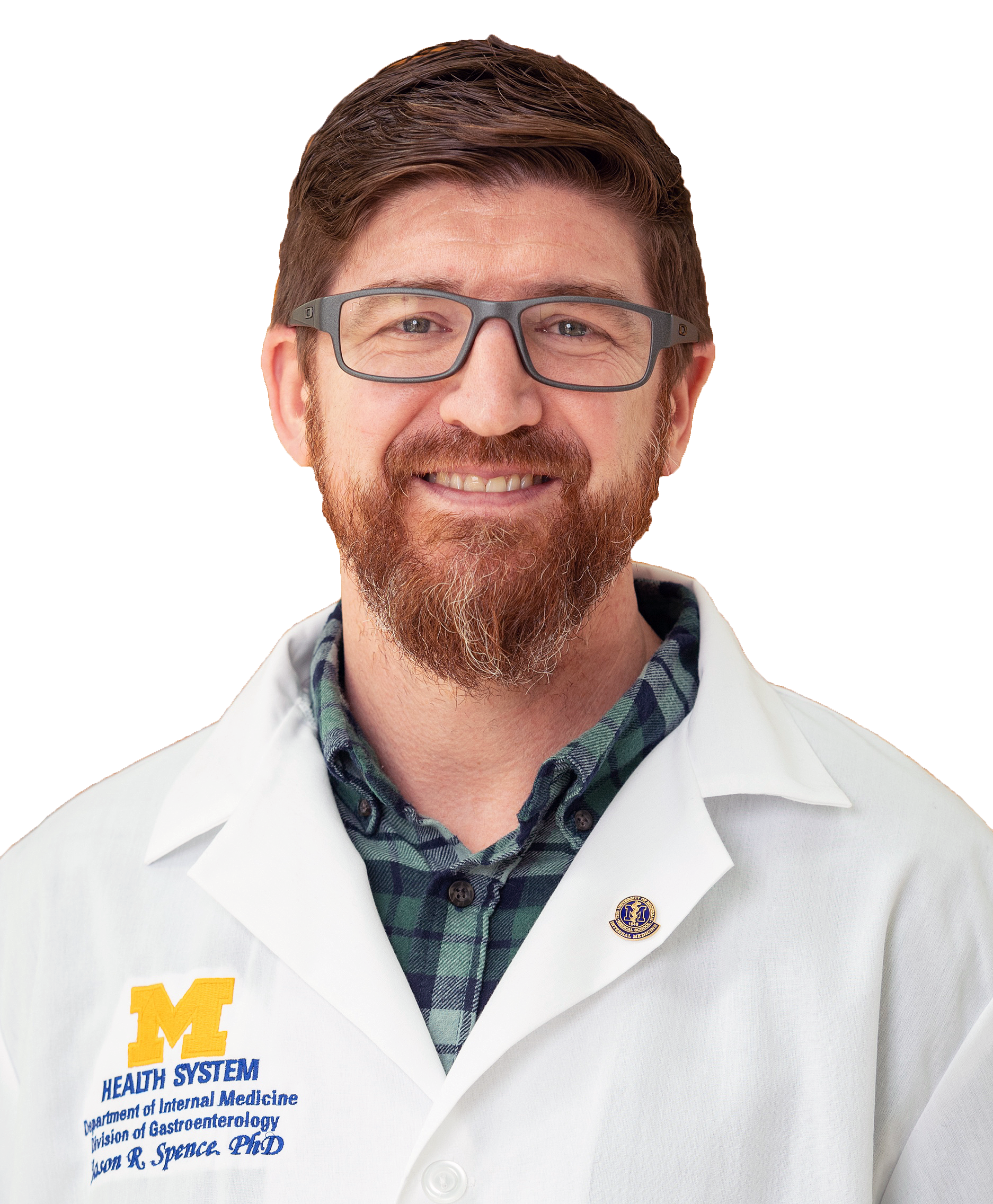 Jason Spence, University of Michigan Medical School
H. Marvin Pollard Professor
Professor of Cell and Developmental Biology, Internal Medicine, Biomedical Engineering“Interrogating Stem Cell Niches During Human Development”
Monday, May 8, 2023, 12:15 p.m. to 1:15 p.m. CDT, 1220 MRBIII
Co-sponsored by the Department of Cell and Developmental BiologyLab website Twitter: @TheSpenceLab
Abstract: The Spence Laboratory uses pluripotent stem cells as building blocks to create multilineage human organs in vitro. These in vitro organ-like structures – called organoids – have transformative potential, from being used as a model system for human development to translational applications in regenerative medicine, cell replacement therapy, and precision medicine. Our vision of turning stem cells into complex tissues is rooted in asking fundamental questions about being human: How do we develop? What goes wrong during disease? How do we heal? In the past, however, interrogating human development has been limited by small and precious tissue samples. Advances in single-cell transcriptomic and spatial imaging technologies, combined with human stem cell and organoid systems have recently flipped this paradigm, opening the door to previously unimaginable insights. Our recent efforts have used these experimental approaches to study human organ development across time. By interrogating human organ development, organ specific stem cells, and stem cell niches, we build hypotheses that can be tested in organoid models, and that can lead to the development of new tools to interrogate human biology. From this work, we have generated rich atlases of human organ development; we have leveraged single cell data to investigate cellular heterogeneity and identified new cell types, and we have identified transcriptional and signaling mechanisms that control organ-specific stem cells in vivo. Our current research is aimed at understanding the function of novel cell types, using human stem cell niche factors to develop life-like organoid models from pluripotent stem cells, and interrogating human disease to inform new strategies for tissue repair and regeneration.
 Volker Dötsch, Institute of Biophysical Chemistry, Goethe University
Professor of Biochemistry"Mechanism of Genetic Quality Control in Germ Cells"
Monday, May 22, 2023 (4:00 pm, 1220 MRB III)
Co-sponsored by the Vanderbilt Center for Structural BiologyAbstract: The survival rate of cancer patients is steadily increasing due to better and more efficient therapies. These advances in cancer therapy, however, create a new and increasing problem since treatment with chemotherapeutic drugs and radiation increases the risk of premature ovarian insufficiency (POI) for female cancer patients. While assisted reproductive technologies can address the problem of infertility, these measures cannot restore the hormonal functions pivotal for women’s health. Although recent advances in whole organ replacement could eventually offer an option to restore long-term hormone function and fertility in the future, a more detailed understanding of the molecular mechanisms of therapy-induced POI could identify targets for pharmacological prevention of POI during gonadotoxic therapies. Loss of the primordial follicle reserve is the most important cause of POI. TAp63a, a member of the p53 family, is the central transcription factor that upregulates the apoptotic program in oocytes. In resting primary oocytes it adopts an inactive state by forming a closed, only dimeric conformation. Detection of DNA damage activates the stress kinases CHk1 and CHk2 which leads to the phosphorylation of TAp63a which itself does not influence the conformation of TAp63a. Instead it recruits another kinase, CK1, which adds four more phosphate groups C-terminally to the CHk2 site. Electrostatic repulsion between the phosphate groups and a stretch of aspartic acids results in the destabilization of the closed dimeric state and the formation of the open and active tetramer. Measuring the phosphorylation kinetics has further shown that the individual CK1 phosphorylation events follow different kinetic regimes which is part of a kinetically encoded safety mechanism that triggers oocytes death only if a certain DNA damage level threshold is reached. This sophisticated system of monitoring the genetic integrity is evolutionary highly conserved but absent in male germ cells.
 John Jumper, DeepMind Technologies
Senior Staff Research Scientist“Highly Accurate Protein Structure Prediction and Its Applications"
Wednesday, August 30, 2023 (4:00 pm, 208 Light Hall)
Co-sponsored by the Center for Applied Artificial Intelligence in Protein DynamicsRead his Nature paper, "Highly accurate protein structure prediction with AlphaFold," here.
View Dr. Jumper's Apex Lecture here.
Abstract: Our work on deep learning, specifically the AlphaFold system, has demonstrated that neural networks are capable of highly accurate modeling of both protein structure and protein-protein interactions. In particular, the system shows a remarkable ability to extract chemical and evolutionary principles from experimental structural data. This computational tool has repeatedly shown the ability to not only predict accurate structures for novel sequences and novel folds but also to do unexpected tasks such as selecting stable protein designs or detecting protein disorder. In this lecture, I will discuss the context of this breakthrough in the machine learning principles, the diverse data and rigorous evaluation environment that enabled it to occur, and the many innovative ways in which the community is using these tools to do new types of science. I will also reflect on some surprising limitations — insensitivity to mutations and the lack of context about the chemical environment of the proteins — and how this may be traced back to the essential features of the training process. Finally, I will conclude with a discussion of some ideas on the future of machine learning in structure biology and how the experimental and computational communities can think about organizing their research and data to enable many more such breakthroughs in the future.
 Ivet Bahar, Stony Brook University
Professor of Biochemistry and Cell Biology
Director, Laufer Center for Physical and Quantitative Biology“Merging Machine Learning Methods and Elastic Network Models for Modeling Protein Dynamics and Function"
Wednesday, September 20, 2023 (4:00 p.m. CDT, 214 Light Hall)
A reception will follow the lecture in the North Lobby of Light Hall.Co-sponsored by the Department of Pharmacology & The Center for Applied Artificial Intelligence in Protein Dynamics.
Abstract: Machine learning (ML)-based approaches have recently shown remarkable
success in predicting protein structures. Progress made in physics-based
computational evaluation of structural dynamics, not only structure, can
also be leveraged if used in conjunction with ML methods. One bottleneck
has been the computing time cost of molecular simulations. However, in
recent years, with the introduction of elastic network models, it is possible to
efficiently evaluate biomolecular properties dependent on dynamics. We
will present recent applications to evaluating the pathogenicity of single
amino acid variants, as well as the impact of deletions on stability.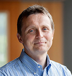 Michael Rapé, University of California, Berkeley
Professor and Head, Division of Molecular Therapeutics
Investigator, Howard Hughes Medical Institute
Dr. K. Peter Hirth Chair of Cancer Biology"Stress Signaling at the Crossroads of Development and Disease"
Friday, September 29, 2023 (12:00 p.m. CDT, 202 Light Hall)Co-sponsored by the Department of Biochemistry. Read more!
Abstract: Human development can withstand many mutational or environmental insults. Key to robust cell fate specification are signaling pathways that detect various stresses, such as nutrient limitation, oxidative damage, or toxin exposure, and in turn instigate reactions that either alleviate or bypass such conditions. How stress responses safeguard tissue formation and homeostasis is still poorly understood. We have recently discovered several stress responses with important roles in cell differentiation, including dimerization quality control, aggregate clearance, or the reductive stress response. These pathways ensure cell homeostasis by controlling processes as diverse as complex formation, protein aggregation, and mitochondrial activity. Using mitochondria as an example, I will describe how cells activate specific stress responses to ensure continuous organellar activity, and I will present our most recent work on active and tightly regulated silencing of these protective signaling networks. Our studies reveal how cells use stress response pathways to adjust organellar function to environmental or developmental needs, and they propose new therapeutic approaches to counteract degenerative disease caused by aberrant mitochondrial function or protein aggregation.
 Katrin Karbstein, University of Florida
Professor, Integrative Structural and Computational Biology"Quality Control During and After Ribosome Assembly"
Tuesday, October 17, 2023 (2:00 p.m. CDT, 1220 MRB III)Co-sponsored by the Department of Biochemistry.
Abstract: Ribosomes are major players in the protein homeostasis network. Not only are they responsible for mRNA selection and protein translation, protein folding begins inside the ribosome, and is influenced by translation. Moreover, because translation is essential for mRNA quality control, the integrity of the mRNA pool depends on the integrity of the ribosome pool. Thus, it is critical that ribosomes are correctly assembled, and maintain full functionality during their life cycle. In our lab we are studying mechanisms for quality control during assembly and translation that allow misassembled ribosomes to be detected and either degraded or repaired
Discovery Science Emerging Scholars Lecture Series
The Discovery Science Emerging Scholars Lectures features young scientists making notable discoveries as postdoctoral fellows or early career faculty.
Emerging Scholars Lectures
-
2024 Speakers
Anthony Venid
 Tuesday, November 19, 4:00 pm, 208 Light Hall. HHMI Hanna Gray Postdoctoral Fellow, Stanford University: “Quality Control in Human Diseases: Autophagy-dependent Mechanisms in Cancer and Neurodegeneration"
In-person only. Co-sponsored by the Department of Cell and Developmental Biology
Nicole Perry-Hauser
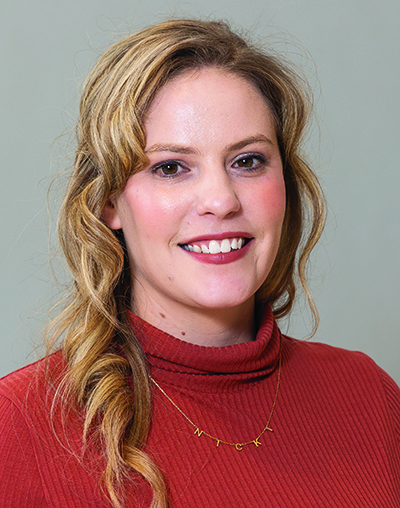 Tuesday, January 23rd, 4:00 pm, 214 Light Hall. Associate Research Scientist, Columbia University, Department of Psychiatry: “Adhesion G Protein-Coupled Receptor Latrophilin-3 as a Novel Target for Modulating Dopaminergic Neurotransmission"
In-person only. Co-sponsored by the Department of Pharmacology. Save-the-date link
Abstract: “Disruption of the adhesion G protein-coupled receptor latrophilin-3 (ADGRL3) results in altered dopaminergic neurotransmission across animal species via an unknown mechanism. Normal dopamine signaling is critical for tasks such as motor behavior, motivation, and working memory, and its dysregulation is implicated in several mental health and neurological disorders. Consequently, ADGRL3 emerges as a promising target for the regulation of dopaminergic neurotransmission. In my research, I use a diverse set of techniques, including biochemical analyses, in-cell assays, and animal models, to investigate how ADGRL3 modulates dopaminergic neurotransmission specifically in the mouse striatum.” Perry-Hauser Flyer
-
2023 speakers
Iris Smith
 Friday, October 20th, 12:00 pm, 202 Light Hall. Iris Smith, Research Instructor, Cleveland Clinic, Lerner Research Institute: "Unraveling PTEN Structural and Functional Dynamic Architecture for Therapeutic Modulation in Cancer and Autism" In-person only. Co-sponsored by the Department of Biochemistry. Save-the-date link
Tumor suppressor gene PTEN, when mutated in the germline, predisposes to PTEN hamartoma tumor syndrome (PHTS), a rare inherited cancer syndrome and, intriguingly, one of the most common causes of autism spectrum disorder (ASD). Currently, it is impossible to predict at the individual level who will develop cancer, ASD, and/or both. Utilizing an integrative structural modeling approach, we demonstrate the structural and conformational effects of clinically relevant PTEN mutations revealing disease-specific molecular features that contribute to autism or cancer and unveil potential allosteric druggable sites to be targeted as a viable and currently unexplored treatment approach for individuals with PHTS. Smith Flyer Smith CV
Jeannette Tenthorey
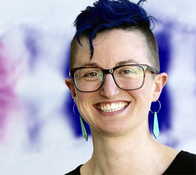 Friday, May 19th, 11:00 am, 214 Light Hall. Jeannette Tenthorey, Assistant Professor and HHMI Hanna Gray Fellow, University of California at San Francisco: "The host strikes back: strategies for evolutionary warfare with viruses" In-person only. Co-sponsored by the Department of Biochemistry. Save-the-date link
Antiviral restriction factors are engaged in evolutionary warfare with viruses, in which viruses mutate to evade restriction factor recognition, and restriction factors then mutate to regain defense. These arms races drive rapid germline evolution of restriction factors, including the retroviral restriction factor TRIM5. Nevertheless, it is unclear how host restriction factors successfully compete in these arms races, given that viruses evolve much faster than their hosts. I explore the hypothesis that antiviral proteins evolve within highly permissive evolutionary landscapes, such that many possible mutations are adaptive in these arms races, thus favoring host participation in such conflicts. Tenthorey CV Tenthorey Flyer @JTenthorey
Celine Riera
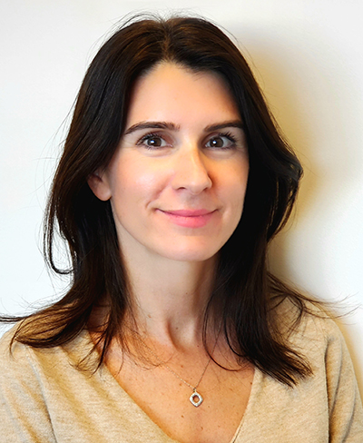 Thursday, March 9, Celine Riera, Assistant Professor, Department of Biomedical Sciences, Cedars-Sinai Medical Center, Los Angeles: "Hypothalamic Sensory Circuits Controlling Adaptive Thermogenesis and Weight Gain" In-person only. Co-sponsored by the Department of Molecular Physiology and Biophysics.
I will share new insights revealing the connection between the perception of the environment and the regulation of energy homeostasis in mouse models. I will describe the neurocircuits controlling energy balance in response to environmental cues including temperature and olfactory stimuli. I will show how these circuits play a role in adaptive thermogenesis and weight gain in animal models of obesity and discuss sexual dimorphism in sensing environmental cues. Riera CV Riera Flyer @celineriera
Hernandez Moura Silva
 Thursday, January 26, Hernandez Moura Silva, Assistant Professor of Biology, MIT, 9:30 am, 206 Preston Research Building: "Perivascular Macrophages in the Intersection of Tissue Health and Disease Pathophysiology" In-person only. Co-sponsored by the Department of Molecular Physiology and Biophysics.
Perivascular macrophages have been described and appear to play a role in tissue physiology, although it remains unclear what are the key features of these cells. We identified a population of perivascular macrophages characterized by their dependence on the transcription factor c-MAF. Furthermore, I observed that c-MAF dependent macrophages actively shape the white adipose tissue vasculature network. Finally, c-MAF ablation in macrophages prevented the development of the early signs of metabolic syndrome upon chronic hypercaloric diet feeding. These data show that tissue resident perivascular macrophages participate actively in the tissue morphogenesis and metabolic syndrome pathophysiology. Silva CV Silva Flyer @hernandezmsilva
-
2022 speakers
2022 Speakers
Sezen Meydan
 Tuesday, November 29, Sezen Meydan, Postdoctoral Fellow, NIH, 4:00 pm, 1220 MRB III: "Multiple Signaling Pathways Link Collided Ribosomes to Cellular Stress Responses" In-person only. Co-sponsored by the Department of Cell and Developmental Biology.
Ribosomes pause upon encountering problems during translation and collide with each other, forming “disome” complexes. Accumulation of disomes is linked to diseases, but how disomes are detected and affect cellular homeostasis is not fully understood. By developing high-throughput sequencing methodologies, we showed that disomes are detected by multiple pathways, including the Integrated Stress Response, which is a major signaling network in eukaryotes induced by environmental stress and pathological conditions. We also found that disome-induced signaling contributes to an antioxidant response during oxidative stress. Our results show that disomes act as signaling platforms to link translation distress into cellular stress responses. Meydan Flyer
Kelsie Eichel
 Tuesday, November 1, Kelsie Eichel, Postdoctoral Fellow, Stanford University, 4:00 pm, 214 Light Hall: "How to Organize a Neuron: From C. elegans to Humans"
Dynamic protein organization within cells underlies fundamental biological processes. This is particularly evident in neurons, which are compartmentalized into distinct axonal and dendritic domains that require unique repertoires of proteins. Loss of polarity in neurons is associated with a wide array of pathologies, underscoring its importance. I identified a conserved and essential mechanism at the axon-dendrite boundary that changes our understanding of how neurons maintain protein compartmentalization over their long lifespan. I found that axonally and dendritically polarized receptors are cleared from the plasma membrane through endocytosis at the axon-dendrite boundary, which prevents receptor diffusion into the inappropriate compartment. Eichel Flyer
Rita Brookheart

Thursday, September 29, Rita Brookheart, Assistant Professor, Washington University School of Medicine, 9:30 am, 206 Preston Research Building: "New Molecular Players Linking Muscle Metabolism and Growth"
Dr. Brookheart’s seminar focuses on her lab’s mechanistic work of how Site-1 Protease (S1P) controls mitochondrial metabolism in skeletal muscle. S1P is a Golgi-resident protease required for the proteolytic cleavage and activation of an array of transcription factors that are required to maintain and restore cellular homeostasis in response to ER stress and disruptions in cellular sterol/lipid levels. Dr. Brookheart’s lab previously identified a patient with a gain-of-function mutation in S1P who exhibited exercise intolerance and abnormal skeletal muscle mitochondrial morphology. Building on this study, her lab defined a previously unknown role for S1P in skeletal muscle metabolism and mass. Brookheart Flyer
Monica Santisteban
 Thursday, April 21, Monica Santisteban, Instructor in Neuroscience, Brain and Mind Research Institute, Weill Cornell Medicine, 9:30 am, 206 Preston Research Building: "Cerebrovascular and Neuroimmune Mechanisms of Cognitive Impairment in Hypertension." Co-sponsored by the Vanderbilt Brain Institute, the Department of Molecular Biology and Biophysics, the Department of Pharmacology, and the Vanderbilt Memory and Alzheimer’s Center.
Dementia affects almost 50 million adults worldwide,and is a major cause of death and disability. Vascular risk factors, such as hypertension and high-dietary salt, have gained importance since up to 50% of clinically diagnosed Alzheimer disease have a mixed pathology at autopsy including cerebrovascular lesions. Thus, elucidating novel mechanisms by which hypertension negatively impacts neurovascular and cognitive function is a necessary step to develop new approaches to protect the brain from hypertension. We have identified two distinct immune mechanisms, involving brain resident perivascular macrophages and circulating IL-17, contributing to cognitive impairment in mouse models of hypertension. Santisteban Flyer
Mary Jane Tsang
 Tuesday, April 12, Mary Jane Tsang, Postdoctoral Fellow at the Whitehead Institute for Biomedical Research, 4:00 pm, 1220 MRB III: “Setting a Timer for Mitosis: How Do Cells Slip Out?” Co-sponsored by the Department of Cell and Developmental Biology.
The Spindle Assembly Checkpoint is the key regulatory pathway that delays anaphase onset until mitotic chromosome segregation defects are corrected. However, in the presence of persistent, unresolvable errors, cells can undergo “mitotic slippage”, exiting mitosis into a tetraploid G1 state and escaping the cell death that results from a prolonged arrest. The molecular logic that allows cells to balance these dueling mitotic arrest and slippage behaviors remains unclear. We have uncovered that human cells modulate their mitotic arrest duration through the presence of conserved, alternative translational isoforms of the essential APC/C co-activator Cdc20, thus providing a new paradigm for controlling cell cycle events. Tsang Flyer
Theresa Loveless
 Thursday, March 10, Theresa Loveless, Associate Project Scientist, Department of Biomedical Engineering, University of California-Irvine, 12:00 pm, 202 Light Hall: "Ordered DNA Writing Enables Lineage Tracing and Analog Recording in Mammalian Cells." Co-sponsored by the Department of Biochemistry.
 Studying multicellular developmental processes can require the non-destructive observation of thousands to billions of cells deep within an animal. DNA recorders address this difficult task by converting transient cellular experiences into changes in the genome that can be sequenced later in high throughput. First, I will present a recently-published DNA recorder that acts primarily by writing new DNA, through the repeated insertion of random nucleotides at a single locus in temporal order, rather than by erasing as previous DNA recording technologies have done. Second, I will share the characterization of a new DNA recording architecture that maintains the strengths of CHYRON – ordered, information-dense recording – but (1) drastically reduces the rate of information loss through off-pathway mutations, (2) is in principle compatible with tens of rounds of recording, and (3) can in principle record at least tens of orthogonal inputs. Loveless Flyer
-
2021 speakers
2021 Speakers
Alex J. Guseman
 Thursday, December 2, Alex J. Guseman, Merck Fellow, University of Pittsburgh Medical School, 9:30 am, 202 Light Hall: "Peering into the Crystallin Ball of the Eye Lens." Co-sponsored by the Department of Molecular Biology and Biophysics.
Cataracts, the opacification of the eye lens, are the leading cause of blindness worldwide. Cataracts form when the crystallin proteins of the eye lens are unable to maintain proteostasis and being to aggregate. As these aggregates grow, the lens opacifies, and visual impairment increases. Despite decades of research on the crystallin proteins, the mechanism of cataract formation is highly debated. Here we demonstrate in vitro that deamidation, a common post translational damage, has minimal impact to crystallin structure and biophysics, and highlight our progress to study crystallins in their native environment, the intact eye lens. Guseman Flyer
Daniel Gonzales
 Tuesday, November 16, Daniel Gonzales, HHMI Hanna Gray Fellow, Purdue University, Weldon School of Biomedical Engineering, 2:00 pm, 1220 MRB III: "Micro- and Nano-scale Technologies for Mapping Sensory-Driven Activity from the Cortical Surface." Co-sponsored by the Vanderbilt Brain Institute.
I will discuss a suite of flexible, transparent electrode technologies for high-density mapping of neural activity from the brain surface in behaving animals. Specifically, we show evidence that surface grids record biomarkers of subcellular activity that travel across the upper cortical layers during sensory stimulation. Thus, when we combine our grids arrays with existing technologies like silicon probes and two-photon imaging, we enable a platform for recording both subcellular dynamics and population-level outputs in awake animals. Gonzales Flyer
Lauren Goins
 Monday, November 8, Lauren Goins, Ph.D., Assistant Project Scientist, UCLA, 12:15 pm CT, 1220 MRB III: "New Paths and Pathways in Developmental Hematopoiesis." Co-sponsored by the Department of Cell and Developmental Biology.
Fundamental questions remain unresolved in the hematopoiesis field such as how hematopoietic stem cells balance proliferation, differentiation, and specialization steps to produce the vast repertoire of blood cell types required for proper immune function. We have tackled this rather complex problem using a multi-pronged approach that combines transcriptomics, genetics, and mutational analysis using the Drosophila hematopoietic system as a model. In the hematopoietic progenitor state, we find that cell-extrinsic signals play a major role in controlling both spatial and temporal aspects of the cell cycle and their differentiation into mature cell types. In particular, we discovered that blood progenitors are maintained in a non-proliferative state in the G2 phase of the cell cycle by Wnt- and hedgehog-dependent signaling pathways that link cell cycle progression to differentiation. Furthermore, our analysis of crystal cells, platelet‐like cells involved in melanization, has uncovered new means by which Musashi and Numb affect canonical and non-canonical Notch signaling. We propose a model of how blood cells integrate various external inputs to determine cell cycle status and differentiation fate. Goins Flyer
Maria Fernanda Forni
 Thursday, October 14, Maria Fernanda Forni, Ph.D., Pew Postdoctoral Fellow, Yale University, 9:30 am CT, 208 Light Hall: "A Central Role for Metabolism in the Skin: Two Tales of How Adipocytes Can Impact Aging and Healing." Co-sponsored by the Department of Molecular Physiology and Biophysics.
Metabolic regulation of tissue homeostasis is one of the most intriguing yet unexplored areas in skin biology. The skin consists of many different tissues and cell types that communicate in a regulated and timely manner to maintain organ homeostasis. As we age, this homeostasis is lost, and with it, the plethora of metabolic reactions that comprise normal homeostasis shift their balance, being perturbed or disrupted. Adipocytes are one of the key players in skin homeostasis regulation. They undergo drastic alterations during the processes of aging and wound healing. In this seminar, I will discuss how these cells are important for whole-body thermal homeostasis and metabolic fitness under conditions of limited energy intake. I will also address how these cells act as metabolic coordinators of immune cell function in the process of wound healing and how aging impacts these processes. Forni Flyer
Sonya Neal
 Friday, April 23, Sonya Neal, Ph.D., Assistant Professor, UC-San Diego, 12:00 pm CT: "Rhomboid Pseudoprotease Dfm1’s Role in ERADicating Multi-Spanning Membrane Substrates."
Nearly one-third of membrane proteins are initially targeted to the endoplasmic reticulum (ER) membrane where they are correctly folded, assembled, and then delivered to their final cellular destinations. In order to prevent the accumulation of misfolded membrane proteins, ER associated degradation (ERAD) moves these clients from the ER membrane to the cytosol; a process known as retrotranslocation. Our recent work in S. cerevisiae has revealed a derlin rhomboid pseudoprotease, Dfm1, is involved in the retrotranslocation of ubiquitinated ERAD membrane substrates. Overall, our work seeks to advance the understanding of rhomboid pseudoproteases in membrane protein quality, a highly conserved process, which is implicated in a plethora of diseases such as cancer, Cystic Fibrosis and neurodegenerative diseases. Neal Flyer
Chrystal Starbird
 Friday, April 2, 12:00 pm CST via Zoom, Chrystal Starbird, Ph.D., Postdoctoral Fellow, Yale University, Department of Pharmacology: "Structural Basis of TAM Receptor Oligomerization." Co-sponsored by the Department of Biochemistry.
Receptor tyrosine kinases are key regulators of a wide range of biological functions and share a single transmembrane helix that links an extracellular domain to an intracellular kinase domain. How a single helix promotes the large diversity of signaling mediated by RTKs has long been a question of interest. Classically, RTKs are considered to be activated by ligand-induced dimerization. However, my work on the TAM subfamily of receptors has shown unique requirements for oligomerization and suggests that our current simple view of receptor activation is insufficient to explain the observed complexity of receptor signaling. Starbird Flyer Get Zoom info here.
Ana Arruda
 Tuesday, March 30, 4:00 pm CST via Zoom. Ana Arruda, Ph.D., Research Specialist, Harvard University School of Public Health, Cell and Developmental Biology. “Interorganelle communication in metabolic health and disease.” Co-sponsored by the Department of Cell and Developmental Biology.
 Metabolically specialized cells display complex spatio-temporal organization of organelles in the tissue context to meet functional demands and adapt to challenges to maintain homeostasis. In this talk, I will discuss how different physiological and pathological nutritional states influence organelle morphology and interorganelle communication in liver and how alterations in these processes relates to the development of metabolic diseases. Arruda Flyer Get Zoom info here.
Daniel Castro
 Wednesday, March 17, 9:00 am CST via Zoom, Daniel Castro, Ph.D., Acting Instructor, University of Washington, Department of Anesthesiology: "Next-Generation Approaches for Investigating Neuropeptidergic Circuits in Reward and Motivation." Co-sponsored by the Department of Pharmacology.
 Here I report that endogenous MOPR regulation of appetitive behavior in mice acts through a specific dorsal raphe to nucleus accumbens (NAc) projection. Select enkephalin-containing NAc ensembles are engaged prior to reward consumption, suggesting that local enkephalin release is the source of endogenous MOPR ligand. Selective modulation of NAc enkephalin neurons and CRISPR-Cas9-mediadated disruption of enkephalin substantiate this finding. Castro Flyer Get Zoom info here.
Florentine Rutaganira
 Thursday, January 21, 4:00 pm CST via Zoom, Florentine Rutaganira, Ph.D., HHMI Hannah H. Gray Fellow, University of California-Berkeley: "Kinase Chemical Genetics in the Closest Living Relatives of Animals."
Although all organisms use signal transduction to respond to external stimuli, the rise of multicellularity necessitated the evolution of signaling pathways to coordinate actions of individual cells into a singular response. As the closest living relatives of animals, choanoflagellates present a unique system to directly study the role of signaling pathways that were previously thought to be restricted to animals. High-throughput screening and activity-based kinase profiling within Salpingoeca rosetta, a choanoflagellate that differentiates into a rosette colony during its dynamic life history, provide evidence for a functional phosphotyrosine signaling repertoire that developed prior to the origin of animals. Rutaganira Flyer Get Zoom info here.
-
2020 speakers
2020 Speakers
Chelsey Spriggs
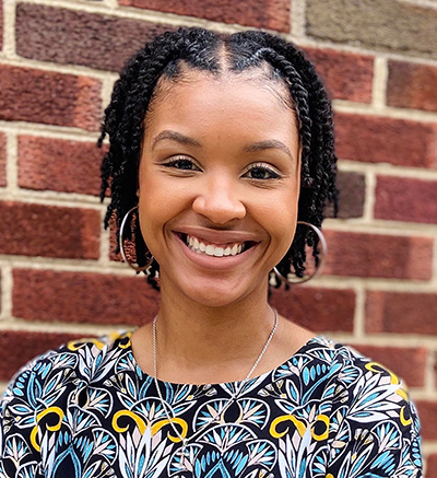 Friday, December 11, 12:00 pm via Zoom, Chelsey Spriggs, Ph.D., Postdoctoral Fellow, University of Michigan Medical Center: "Viral hijacking of host molecular motors to promote nuclear entry." Co-sponsored by the Department of Biochemistry.
 During entry, most DNA viruses must navigate the crowded cellular environment to reach the nucleus where transcription and replication of the viral genome occur. How polyomavirus (PyV), a small DNA tumor virus, accomplishes this feat is not well-understood. We reported that cytoplasmic dynein motor activity is required to disassemble PyV once in the cytosol and to target the disassembled virus to the nucleus. However, the precise mechanism underlying these dynein-dependent steps remain unclear. Investigating the role of dynein cargo adaptor proteins in this process, we have discovered an unexpected and novel function of the bicaudal-D adaptors in virus infection. Spriggs Flyer Get Zoom info here.
Christina Termini
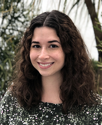 Tuesday, December 8, 4:00 pm via Zoom, Christina Termini, Ph.D., University of California President’s Postdoctoral Fellow, UCLA: “Leveraging proteoglycans for hematopoietic stem cell regeneration.” Co-sponsored by the Department of Cell and Developmental Biology.
 Although hematopoietic stem cells (HSCs) comprise less than 0.01% of the adult bone marrow, this rare cell population is capable of supporting the hematopoietic needs of an organism throughout their lifetime. However, when HSCs are unable to adapt to hematopoietic stress, life-threatening complications can arise. As such, it is essential we elucidate the basic regulatory mechanisms that underscore HSC regeneration. My postdoctoral research identified a novel surface marker that both identifies and regulates HSCs through parallel mechanisms. This surface marker is from the heparan sulfate family of proteoglycans and can be used to enrich for long-term HSCs capable of enhanced hematopoietic engraftment, expansion and self-renewal ability. Loss of function analyses determined that this heparan sulfate proteoglycan regulates HSC maintenance and myelopoiesis through alterations in cell cycling. Additional work analyzing how proteoglycans can be utilized to accelerate hematopoietic recovery after myelosuppression will be presented. Termini Flyer Get Zoom info here.
Cornelius Taabazuing
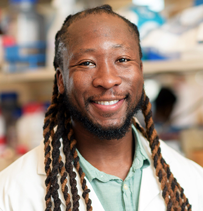 Thursday, November 19, 9:30 am via Zoom, Cornelius Taabazuing, Ph.D., Postdoctoral Fellow, Memorial Sloan-Kettering Cancer Center: "Investigating the molecular regulation and function of death signaling complexes." Co-sponsored by the Department of Molecular Physiology & Biophysics.
The cellular response to danger signals such as pathogens or DNA damage is important for survival and many diseases. The response is usually mediated by large multiprotein death signaling complexes that can either eliminate the damaged cells or promote homeostatic restoration pathways. However, the specific inputs that initiate the death signaling complex formation as well as the downstream signaling pathways activated by these complexes are not well understood. Here, we demonstrate how small molecule inhibitors of the serine peptidases, DPP8/9, induce formation of the CARD8 and NLRP1 inflammasomes, which activate caspase-1 to cause an immunostimulatory type of death called pyroptosis. Taabazuing Flyer Get Zoom info here.
Melanie McReynolds
 Thursday, October 29, 9:30 am via Zoom, Melanie McReynolds, Ph.D., HHMI Hanna H. Gray Fellow, Princeton University, "NAD+ flux is maintained in aged mice." Co-sponsored by the Department of Molecular Physiology & Biophysics.
NAD+ is an essential coenzyme found in all living cells. NAD+ concentrations decline during aging, but whether this reflects impaired production or accelerated consumption remains unclear. Here we employed isotope tracing and mass spectrometry to probe NAD+ metabolism across tissues in aged mice. In 25-month-old mice, we observe modest tissue NAD+ depletion without significant changes in circulating NAD+ precursors. Isotope tracing showed unimpaired synthesis of circulating nicotinamide from tryptophan, and maintained flux of circulating nicotinamide into tissue NAD+ pools. Although absolute NAD+ biosynthetic flux was maintained in most tissues of aged mice, fractional tissue NAD+ labeling from infused labeled nicotinamide was modestly accelerated. Long-term calorie restriction partially mitigated age-associated NAD+ decline despite decreasing NAD+ synthesis. Thus, age-related decline in NAD+ is driven by increased NAD+ consumer activity rather than impaired production. McReynolds Flyer
Antentor “A.J.” Hinton, Jr.
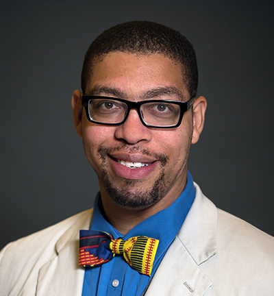 Thursday, March 5, Antentor “A.J.” Hinton, Jr., Ph.D., 9:30 am, 206 Preston Research Building: "OPA-1 Deficiency in Skeletal Muscle Increases Mitochondria ER Contact Formation Through an ATF-4 Dependent Mechanism."
Dr. Hinton, Jr. is a Postdoctoral Research Fellow in the Department of Internal Medicine, University of Iowa. In the laboratory of E. Dale Abel we have found that reduction in OPA1 (Optic Atrophy 1, a mitochondrial dynamin-like GTPase) expression in skeletal muscle results in endoplasmic reticulum (ER) stress response and FGF21 secretion. We predicted that there was an association between the loss of OPA1, an increase in ER-Mitochondrial Contact sites, and prevalence of diabetes. Mitochondrial endoplasmic reticulum contact sites (MERCs) are specialized membranes that are enriched with specific proteins believed to be important for calcium flux, lipid transfer, and mitochondria morphology. This observation provides a plausible mechanism linking altered mitochondrial dynamics with the activation of the ER stress response pathway and may give novel insight into how some patients develop insulin resistance. We propose to use this information to provide the foundation for future research that will lead to the discovery of more effective biomarkers that identify insulin resistance in skeletal muscle. Hinton Flyer
-
2019 speakers
2019 Speakers
Chantell Evans
 Tuesday, December 3, Chantell Evans, 4:00 pm, 206 Preston Research Building: "The Role of Optineurin in Neuronal Mitophagy."
Dr. Evans is an HHMI Hannah H. Gray Postdoctoral Fellow at the University of Pennsylvania. Mitophagy, the selective removal of damaged mitochondria, is thought to be critical to maintain neuronal homeostasis. Mutations in proteins implicated in mitophagy, including PINK1, Parkin, OPTN, and TBK1, cause Parkinson’s disease or ALS, suggesting defective mitochondrial turnover contributes to neurodegeneration. To test this hypothesis, we used mild oxidative stress to induce low levels of mitochondrial damage in hippocampal neurons. We observed the sequential recruitment of Parkin, TBK1, and OPTN to depolarized mitochondria followed by their sequestration into autophagosomes, and determined this pathway was compartmentally restricted to the soma. Further, acidification of mitophagosomes was remarkably slow in neurons and overall was a rate-limiting step in the mitophagy pathway. Expression of an ALS-linked OPTN mutation disrupted the integrity of the mitochondrial network and this effect was exacerbated by oxidative stress. We propose that the slow kinetics of mitophagy enhance neuronal susceptibility to disease-associated mutations in the pathway, leading to neurodegeneration. Evans Flyer
Reginald D. Cannady
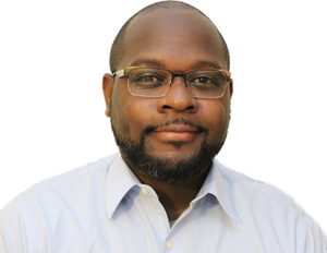 Tuesday, November 12, Reginald D. Cannady, 4:00 pm, 512 Light Hall: “Probing Novel Targets to Reduce Heavy Drinking in Models of Alcohol Use Disorder."
Dr. Cannady is a Postdoctoral Research Fellow at the Medical University of South Carolina. Individuals with alcohol use disorder (AUD) show an inability to regulate alcohol consumption and seeking behavior. Although, there are FDA‐approved medications for AUD, they have limited efficacy. My work examines neuroadaptations caused by chronic alcohol experience and focuses on identifying neuronal targets to reduce heavy drinking using several rodent models of alcohol addiction. This presentation will discuss the validation of KV3 potassium channels as a potential therapeutic target to reduce alcohol consumption in preclinical models of alcohol dependence. Cannady Flyer
Sabena Conley
 Tuesday, October 22, Sabena Conley, 4:00 pm, 512 Light Hall: "Obesity-Induced Mesenchymal Stem Cell Senescence and Dysfunction."
Dr. Conley is a Research Fellow at the Mayo Clinic College of Medicine, Rochester, Minnesota. Endogenous mesenchymal stem cells (MSC) mediate tissue repair, but obesity might potentially impair their regenerative potency. The adipose tissue microenvironment in obesity stimulates cellular senescence (CS), an irreversible cell cycle arrest that prompts an inflammatory, senescent-associated secretory phenotype (SASP). To test the hypothesis that obesity induces CS and dysfunction in MSC from obese patients, we harvested and evaluated MSC from fat tissue collected from obese and non-obese patients undergoing surgery at Mayo Clinic. Obese-MSC demonstrated increased CS and SASP and lower proliferation than non-obese-MSC. Therefore, human obesity induces senescence in adipose tissue-derived MSC, which might interfere with their repair capacity. Conley Flyer
Timothy Downing
 Thursday, April 25, Timothy Downing, 4:00 pm, 512 Light Hall: "Synthetic Genome Regulation for Cell and Tissue Engineering."
Dr. Downing is an Assistant Professor in the Department of Biomedical Engineering, University of California, Irvine. The Downing Lab is interested in understanding how the chemical and biophysical microenvironment influences adult cell behavior and phenotype through epigenetic gene regulatory mechanisms. We hope to use this information in the design of next-generation biomaterials. This presentation will describe how specific cues (e.g., topography and matrix stiffness) within cellular microenvironments can influence epigenetic state and facilitate the reprogramming of somatic cells back to pluripotency. Web Downing Flyer
Luis Natividad
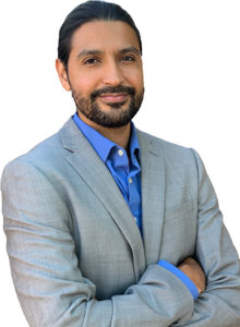 Tuesday, March 5, Luis Natividad, 4:00 pm, 206 Preston Research Building: "Impaired Endocannabinoid Signaling in Stress and Addiction". Co-sponsored by the Vanderbilt Center for Addiction Research.
Dr. Natividad is a Senior Research Associate, The Scripps Research Institute, La Jolla. Abstract: "Heavy alcohol consumption induces long-term problems with stress and anxiety, and is common among dependent individuals who are co-diagnosed with mood disorders. As endocanna-binoids (e.g., N-arachidonoylethanolamine and 2-arachidonoylglycerol) provide an important mechanism of inhibitory constraint in the regulation of stress circuits, Dr. Natividad will elaborate more on the premise of dysregulated endocannabinoid signaling influenced by chronic alcohol exposure relative to observations in a genetic model of “innate alcohol dependence” within the central nucleus of the amygdala." Web Natividad Flyer
Stacey Finley
 Thursday, February 7, Stacey Finley, 4:00 pm, 512 Light Hall: "Systems Biology Approaches to Predict the Dynamics of Biochemical Networks in Cancer."
Dr. Finley is an Assistant Professor at the University of Southern California. Abstract: "Systems biology approaches, including computational models, provide a framework to optimize effective therapeutic strategies for cancer. My research group develops mechanistic models of biochemical networks in cancer to study cancer immunotherapy, tumor angiogenesis, and cancer metabolism. Our models provide insight into the dynamics of the biochemical pathways in cancer and enable the development of novel therapeutics." Website Finley Flyer
-
2018 speakers
2018 Speakers
Angelina Hernandez-Carretero
 Thursday, December 6, Angelina Hernandez-Carretero, 9:30 am, 206 Preston Research Building: "Novel Regulators of Obesity-Induced Insulin Resistance and Diabetes."
Dr. Hernandez-Carretero is a Staff Scientist at the City of Hope Beckman Research Institute. "Further investigation is needed to understand the molecular players involved in obesity-induced insulin resistance. We have utilized a dietary switch mouse model to perform transcriptomics of the skeletal muscle, and compared these findings with RNA-seq of human obese- diabetic muscle. This multispecies approach identified key genes that tracked with the insulin resistant state in both mouse and human muscle, including Cysteine and glycine-rich protein 3 (Csrp3). Csrp3 expression is decreased in obese-insulin resistant muscle and plays an important role in insulin-stimulated glucose uptake." Flyer
Luisa Escobar-Hoyos
 Tuesday, November 13, Luisa Escobar-Hoyos, 4:00 pm, 206 Preston Research Building: “Targeting RAS and mutant p53: Discovery of RNA splicing as a therapeutic vulnerability in pancreatic cancer.”
Dr. Escobar-Hoyos is a Postdoctoral Research Fellow at Memorial Sloan Kettering Cancer Center and Research Assistant Professor at Stony Brook University. "We recently discovered a novel mechanism of cooperation between the two most common oncogenes in pancreatic cancer, oncogenic RAS and neomorphic mutant p53, uncovering a potential therapeutic opportunity to target tumors that bear these mutations. Specifically, we found that mutant p53 causes aberrant splicing of GAP proteins, the negative regulators of RAS, resulting in expression of inactive GAP proteins (polyC GAPs), and ultimately promoting oncogenic RAS signaling. In addition, we identified that pancreatic tumors in mouse models depend on expression of polyC GAPs and splicing machinery proteins, as genetic and chemical inhibition of these proteins caused decrease in tumor growth, number of metastases and tripled the survival time of animals. These studies identified these proteins as new targets for tumors with neomorphic p53 and oncogenic KRAS. We expect to identify novel and specific dependencies of PDAC cells by studying and targeting specific alternatively spliced products and/or manipulating the function of splicing factors in the background of multiple forms of mutated TP53, to provide the foundation for future research that will lead to the development of more effective approaches to treat PDAC patients, improving their survival and quality of life." Flyer
Leonard Alfredo Harris
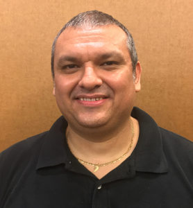 Wednesday, October 10, Leonard Alfredo Harris, Vanderbilt University, 4:00 pm, 512 Light Hall: "Bet Hedging as a Survival Strategy in Complex Biological Systems and Cancer.”
 Cells are complex, dynamic systems capable of initiating internal biochemical programs and adjusting their behavior in response to microenvironmental signals and stressors. Under the general term “phenotypic plasticity,” this phenomenon underlies important biological processes such as stem cell differentiation and epithelial-to-mesenchymal transitions. In bacteria, isogenic cell populations are known to exploit plasticity by phenotypically diversifying to increase their chances of survival to catastrophic external challenges, a strategy known as “bet hedging.” Recent evidence suggests that cancer cells may employ a similar strategy to survive the initial onslaught of anticancer therapeutics. Understanding the complex biochemical networks that underlie signal propagation and cell fate decisions is thus critical for improving treatments of various human diseases, including cancer. Here, I describe the biochemical basis for phenotypic plasticity within the framework of “Waddington landscapes,” present evidence for its role in anticancer drug resistance in non-small cell lung cancer and melanoma populations treated with targeted drugs, and discuss initial work toward constructing a detailed computational model of the biochemical machinery underlying cellular responses to external perturbations. Flyer
Ishmail Abdus-Saboor
 Thursday, May 3: Ishmail Abdus-Saboor, University of Pennsylvania, 4:00 pm, 512 Light Hall: "Genetic Interrogation of Neural Circuit Mechanisms for Pain."
The nervous system is exquisitely tuned to mount the appropriate behavioral response to somatosensory stimuli ranging from a gentle caress to a harsh mechanical insult. How our nervous systems encode this information, from the level of sensory neuron activation in the skin up towards the central nervous system, in both normal and diseased states, remains enigmatic. We are working to uncover the mechanisms governing sensory encoding of touch, itch, and pain. Using optogenetics, quantitative analysis of animal behavior, and in vivo calcium imaging we have 1) determined how a population of pain-sensing neurons have unique morphological and physiological outputs depending upon body location, and 2) developed a new behavioral platform using high-speed videography, statistics, and machine learning to distinguish between innocuous versus painful behavior responses. Flyer Abdus-Saboor Lab
Gustavo Silva
 Thursday, March 1: Gustavo Silva, Duke University, 4:00 pm, 406 Preston Research Building: "K63 ubiquitin and the regulation of translation in response to oxidative stress."
Ubiquitin is a prominent post-translational modification, which signals extensively beyond protein degradation. In this talk, Dr. Silva will present his research on how ubiquitin modifies ribosomes and controls cell resistance to stress. Flyer
Dionna Williams
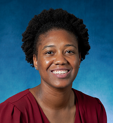 Thursday, February 1: Dionna Williams, Johns Hopkins University, 4:00 pm, 512 Light Hall: “Beyond GPCR Recycling: B-Arrestin as a Neuroprotective Modulator of Innate Immune Responses."
Dr. Williams' talk will focus on the organ-specific, immunomodulatory roles of B-arrestin that serve to suppress chronic immune activation during viral infection of the brain. Flyer
-
2017 speakers
2017 Speakers
Arion Kennedy
 Thursday, December 14: Arion Kennedy, Vanderbilt University, 4:00 pm, 206 Preston Research Building: "CD8+ T Cells Regulate Liver Injury In Obesity-Related Nonalcoholic Fatty Liver Disease."
The incidence of NAFLD has increased in Western countries due to the prevalence of obesity. Current interests are aimed at identifying the type and function of immune cells that infiltrate the liver and key factors responsible for mediating their recruitment and activation in obesity-associated NAFLD. Dr. Kennedy's talk will focus on the role of CD8+ T cells in the development of obesity- associated NAFLD. Flyer
Avery Posey, Jr.
 Thursday, November 16: Avery Posey, Jr., University of Pennsylvania, 12:00 pm, 898 Preston Research Building: "Accelerating CAR T Cells from the Model T to Driverless."
Dr. Posey will discuss engineered T cell therapies developed to treat cancer, including progress and imitations of translating the success of leukemia treatments to solid tumor treatments. Flyer
Sade Spencer
 October 5: Sade Spencer, Medical University of South Carolina, 4:00 pm, 206 Preston Research Building: "Dopaminergic Regulation of Relapse-Dependent Glutamatergic Plasticity."
Dr. Spencer will present her modified cue-cocaine relapse model and the alterations in transient synaptic plasticity associated with relapse observed in the nucleus accumbens. She will also present newer data pertaining to the relationship between dopamine and glutamate during relapse, and how this impacts synaptic function. Flyer
Stephanie Correa
 April 13: Stephanie Correa, UCLA, 4:00 pm, 206 Preston Research Building: "Sex-Specific Neural Regulation of Energy Balance."
The focus of Dr. Correa’s lab is understanding how reproductive hormones affect metabolic health and disease. Flyer Photos
Rene Raphemot
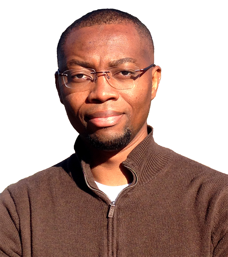 March 16: Rene Raphemot, Duke University, 4:00 pm, 206 Preston Research Building: "A Genomic Screen Reveals New Host Factors Critical to Liver-Stage Malaria."
Dr. Raphemot studies host-parasite interactions during the liver-stage of malaria infection. His goal is to understand host-related factors that are exploited by the parasite. Flyer Reporter Article Photos
Lab-to-Table Conversations
How does fundamental biomedical discovery and its applications connect with your day-to-day life? How does research impact and inform society? Through a new monthly event series called Lab-to-Table Conversations, the Vanderbilt University School of Medicine Basic Sciences looks to explore these questions. View all of our upcoming and previous Lab-to-Table conversations here!
Lab-to-Table Playlist
Lab-to-Table Conversation: Biotech Entrepreneurship: Stories & Strategies
Lab-to-Table Conversations: Picturing Progress: Representation in Scientific Art
Lab-to-Table Conversations: Beyond Addiction: Therapeutic Developments and Societal Impact
Genetic Information: Balancing Ethics and Impact
Jason Isbell: Songs and Sobriety
Meeting the Challenge: Neuroscience Drug Discovery with the Warren Center
A Promising Start to Ending Coronaviruses Webinar
Depression and the Brain: A Paradigm Shift in Psychiatry and Neuroscience
From Inclusion to Equity: The Story of Black Biomedical Scientists panel discussion 2/15/21
On the Cutting Edge: Research Toward a Cure for Colorectal Cancer
Addiction, Sobriety and Art in the Time of COVID-19
Vanderbilt Center for Addiction Research — a conversation with Langhorne Slim
How the Brain Remembers
Lab-to-Table Conversations: Origins of Life
