
Elongating Microvilli in the Intestine
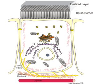
Enterocytes, the epithelial cells lining the lumen of the intestine, develop a dense lawn of microvilli (MV) on their apical surface as they differentiate from stem cells in the intestinal crypt. We know that MV are membrane protrusions supported by a bundle of actin filaments, but we understand very little about how these structures elongate during enterocyte differentiation. This led Vanderbilt Basic Sciences investigator Matthew Tyska and his laboratory to explore the role of IRTKS (insulin receptor tyrosine kinase substrate) in MV formation. IRTKS is a member of the I-BAR (inverse-bin-amphiphysin-Rvs) protein family, characterized by an I-BAR domain, a three-helix bundle that dimerizes and adopts a curved shape that closely matches the membrane curvature of the MV tip. I-BAR proteins also possess an actin-binding Wiskott-Aldrich syndrome protein homology 2 (WH2) motif and an SH3 domain that enables them to interact with other proteins. IRTKS, the only I-BAR protein expressed in the small intestine, is the target of the EspFuvirulence factor secreted by enterohemorrahagic Escherichia coli(EHEC), which damages the intestinal wall by destroying MV. The Tyska lab began their studies by exploring the localization of IRTKS in small intestine organoids, which recapitulate enterocyte differentiation. They found IRTKS to be enriched at the apical surface, particularly in differentiating crypt cells, and concentrated at the tips of MV. IRTKS localization was similar in W4 cells, an epithelial cell line that can be induced to differentiate, forming MV. shRNA-mediated knockdown of IRTKS in M4 cells led to a reduction in both the number and length of developing MV. Expression of selected loss-of-function mutant IRTKS proteins demonstrated that the I-BAR domain was required for targeting to the MV tip, whereas the SH3 domain contributed to but was not totally necessary for tip-targeting, and the WH2 domain was not required. However, overexpression of SH3 or WH2 loss-of-function mutants suppressed MV elongation – presumably by acting via a dominant negative mechanism – indicating that both of these domains play a role in the process. The investigators noted that EspFufrom EHEC, which binds tightly to the IRTKS SH3 domain, exhibits strong homology to EPS8, an endogenous actin bundling and capping protein. This led them to hypothesize that EPS8 might be an important IRTKS interacting partner. Consistently, they found that the two proteins colocalize in MV tips, and that FLAG-tagged EPS8 is pulled down together with EGFP-tagged IRTKS, confirming an interaction between the two proteins. shRNA-mediated knockdown of EPS8 results in similar effects on MV elongation as seen with knockdown of IRTKS, and co-expression of the two proteins in unrelated cells increases the formation of filopodia, a process requiring actin bundle formation and membrane protrusion. The findings support the hypothesis that IRTKS promotes MV elongation by associating with the MV tip via its I-BAR domain and facilitating actin bundling directly via its WH2 domain and indirectly via its association with EPS8. The work is published in the journal Current Biology[M. M. Postema, et al. (2018) Curr. Biol., published online September 6, DOI:10.1016/j.cub.2018.07.022].