By Cayetana Arnaiz Yépez

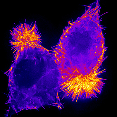
Cells along our intestinal tract are responsible for absorbing nutrients and acting as a barrier to pathogenic microorganisms. During differentiation, these cells, known as enterocytes, develop a specialized structure in their apical surface known as the brush border, which is made up of an array of actin-based protrusions called microvilli. This feature allows them to amplify the membrane surface area and therefore increase their functional capacity. A recent study from the lab of Matt Tyska (Cell and Developmental Biology) parsed out the necessary components for the regulation of microvilli growth in enterocytes. The paper was published in Current Biology.
Despite our vast knowledge about the components present at the brush border, the question of how these components assemble to build and maintain the structure is still unclear. The study, led by postdoctoral fellow James J. Faust, investigated how monomeric actin (G-actin) availability is regulated during microvilli assembly. The work showed that the rate of brush border assembly is limited by the size of the G-actin pool during enterocyte differentiation.
To understand the mechanistic details of this cellular process, they used a mouse model and a human intestinal epithelial cell model. They first tested how changes in the G-actin pool affected brush border formation. To do this, they treated mice with CK-666, a small molecule inhibitor of Arp2/3, a protein that is responsible for the creation of new actin filaments that ‘branch out’ from existing filaments. Inhibiting this protein releases G-actin from branched actin networks, thereby increasing the pool of free G-actin subunits available for the construction of the linear molecules that are observed in microvilli. The results showed that enterocytes from treated mice had longer microvilli than enterocytes from control mice, likely due to increased G-actin availability in the treated mice. The researchers replicated their results in vitro by treating human intestinal epithelial cells with CK-666, but also noted that microvilli assembled faster in treated cells.
To test the idea that accelerated microvilli assembly is due to an increase in the G-actin pool, they used another drug, called latrunculin A, known to sequester actin monomers, to counter the effects of Arp2/3 inhibition. When cells were treated with both CK-666 and latrunculin A, both the length of their microvilli and the brush border formation rates went returned to control levels. Interestingly, when CK-666-treated cells were exposed to increasing concentrations of latrunculin A, the percentage of cells with brush borders significantly decreased, indicating that brush border assembly is likely driven by G-actin availability.
Finally, the authors looked at the actin-binding protein profilin-1 since recent studies indicate that it acts as an Arp2/3 antagonist in some contexts. To examine its functional role within the context of microvilli and the brush border, they performed knockdowns of profilin-1 in cells, which significantly inhibited brush border formation. When they treated these profilin-1-depleted cells with CK-666, there was a reduction of microvilli lengths and no acceleration in brush border formation Therefore, they concluded that removing profilin-1 from cells prevented the protein from directing the G-actin that is released from Arp2/3-based networks to the growing microvilli of these cells.
This paper suggests that G-actin availability and allocation within the cell are crucial for brush border formation. Even though the study focused on the assembly of the brush border on enterocytes, this G-actin pool regulation mechanism could also be important in other cellular contexts that depend on actin-network remodeling, such as cell migration and cell division.