By Jaime Jensen
This article originally appeared in the Nature Research Microbiology Community blog.
The bacterium Clostridioides* difficile (C. diff) has been called many things: a superbug, a public health menace, an urgent concern, a notable threat. C. diff infection (CDI) is frequently nosocomial and results from major disruption of the gut’s normal flora – a state often induced through antibiotic usage. Public health officials have also noticed a rise in community-acquired CDI in recent years. Although the major virulence factors of CDI are toxin A (TcdA) and toxin B (TcdB), a third toxin, C. difficile transferase (CDT), has risen in prominence, as its presence during infection has been associated with more severe forms of disease.
I, along with other members of the lab of Borden Lacy (Pathology, Microbiology & Immunology), recently published a study in Nature Microbiology detailing our recent work on CDT. Through the use of cryo-EM, we identify four CDT structural states, which provided insights into its functioning and helped us identify a previously unknown CDT domain.
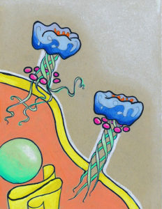
CDT is composed of two proteins, CDTa and CDTb, and is therefore considered a binary toxin. Once it is secreted from C. diff, CDTb binds to host colonic epithelial cells through the lipolysis-stimulated lipoprotein receptor (LSR) and oligomerizes to form a heptamer capable of recruiting CDTa. After the complex is internalized by the colonic cells through endocytosis, a series of intricate conformational changes converts the CDTb heptamer from a prepore to a membrane-spanning pore that completely traverses the lipid bilayer and that allows for the translocation of CDTa into the host cytosol. CDTa then catalyzes ADP-ribosylation of cellular actin, which disrupts the cytoskeleton and ultimately leads to better adherence of C. diff in the colon.
What began as a straightforward approach to structurally characterize CDTb led to a detailed description of CDTb pore formation, provided several clues toward a mechanistic explanation for CDT intoxication, and truly highlighted the immense potential of cryo-electron microscopy (EM).
An observation that in vitro trypsinization yielded oligomerized CDTb, which could be visualized as a dimer of heptamers via negative stain EM, made CDTb an ideal candidate for cryo-EM experiments. What we initially thought was a homogenous population of CDTb particles after size exclusion chromatography and negative stain EM turned out to contain multiple species of CDTb oligomers, which could not be separated despite our best efforts. Included within this heterogeneous group were CDTb monomers and dimers, that, due to limited available views of these smaller particles, only returned low resolution reconstructions, plus a “short” and a “long” form of the double heptamer. Through 3D reconstructions at 3.7-3.9 Å resolution, we showed that the short form contained one heptamer in the pore state, and one in the (non-inserted) prepore state, while the long form contained two very similar heptamers in an intermediary state wherein the beta-barrel is only partially formed. Thus, from a single sample, we successfully isolated three distinct states of CDTb from the transition of prepore to pore.
A second experiment to obtain the structure of CDTb bound to its host cell receptor, LSR, did not show LSR and an expected CDTb domain, but did reveal a fourth “pre-insertion” state of CDTb. By solving the structure of CDTb, we also identified a previously uncharacterized domain that is capable of binding certain glycans and is not present in the structurally related protective antigen from the Bacillus anthracis anthrax toxin.
This work generated several unexpected results, some of which are highlighted above, but one of the most intriguing is that the four CDTb structural states were all observed at pH 8.0. Canonically, the transition to the pore state includes structural changes triggered by the acidic environment of the endosome. However, we have demonstrated that at least some of these extensive structural rearrangements may proceed near physiological pH. Further experiments are required to evaluate the influence of pH on pore formation and cargo translocation, but these current studies considerably enhance our understanding of CDT function and will inform future efforts to develop new CDI therapeutics that specifically neutralize CDT.
This research was facilitated by the VUMC Flow Cytometry Shared Resource and by funding from the United States Department of Veterans Affair, the National Institutes of Health, and Vanderbilt University.
*Formerly Clostridium