By Hillary Layden
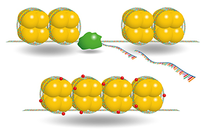
Most human cells contain roughly 6.5 feet of DNA, which must be tightly compacted to fit within the nucleus. Cells compact DNA by wrapping it around proteins called histones, forming a DNA-protein complex called chromatin. “Closed” chromatin is tightly compacted and cannot interact with other proteins, but it can be “opened” by dislodging histones and making the DNA accessible. This process of changing chromatin accessibility is dynamic and essential for gene expression.
Gene expression can also be impacted by the chemical modification of DNA bases, the building blocks of DNA. The most common chemical modification of DNA is the addition of a methyl group, a single carbon with three hydrogens, to cytosine, one of the four DNA bases. Low levels of DNA methylation correlate with active gene expression, and high levels correlate with no expression.
Multiple studies underscore the importance of chromatin accessibility and DNA methylation in development and disease progression. Both features are incredibly important for establishing cell type during development, as each cell type must only express a certain subset of the genes contained in the genome, and are frequently altered by diseases such as cancer. However, a genome-wide understanding of the relationship between chromatin accessibility and DNA methylation has been delayed by a lack of sufficient molecular tools.
A recent study led by Kelly Barnett, a Ph.D. candidate in the lab of Emily Hodges (Biochemistry), describes a new technique, ATAC-Me, which measures chromatin accessibility and DNA methylation within the same DNA molecules. The technique, published in Molecular Cell, is the first of its kind to provide the resolution and coverage required to study DNA methylation and chromatin accessibility together, and is an improvement over previous methods, which could only look at populations of cells and made it difficult to determine if methylation and accessibility in fact coexisted within the same cell. ATAC-Me combines two widely used techniques: an assay that isolates open chromatin and a method that converts unmethylated cytosines to uracil, a base typically found in RNA. Paired with DNA sequencing, ATAC-Me identifies methylated regions within open chromatin.
The development of ATAC-Me was prompted by the observation that enhancers appeared to be regions of overlapping open chromatin and methylated DNA. Enhancers are regulatory regions within chromatin that increase the likelihood that a gene will be transcribed, and are critical for the activation of cell-type-specific genes during development and differentiation. The Hodges group believed that the regions of methylated open chromatin could be the intermediate stages of enhancer activation.
With ATAC-me, the authors definitively showed that areas of open and methylated chromatin exist within the same DNA molecules. They also showed that enhancer regions open and increase gene expression before methylation is removed, providing new insight into the biology of enhancer activation. ATAC-me will greatly increase our understanding of how chromatin accessibility and DNA methylation control these processes under normal circumstances and in disease states.
This study was funded by the National Institutes of Health.