Colon
-

Grant spurs effort to map biology of Crohn’s disease
Nov. 20, 2019, 4:14 PM by Bill Snyder Vanderbilt University Medical Center (VUMC) has been awarded a three-year, $3 million grant from The Leona M. and Harry B. Helmsley Charitable Trust to map — in unprecedented detail — the biology of Crohn’s disease. Under the terms of the award, which… Read MoreNov. 21, 2019
-
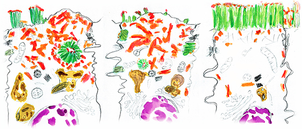
Help! Microvilli trapped inside cells!
By Colbie Chinowsky Drawing of two enterocytes representing Microvillus Inclusion Disease (left and center) and a healthy enterocyte with its microvilli on its apical side. Adapted with permission from Vogel, GF, Janecke, AR, Krainer, IM, Gutleben, K, Witting, B, Mitton, SG, Mansour, S, Ballauff, A, Roland, JT, Engevik, AC, Cutz,… Read MoreOct. 28, 2019
-

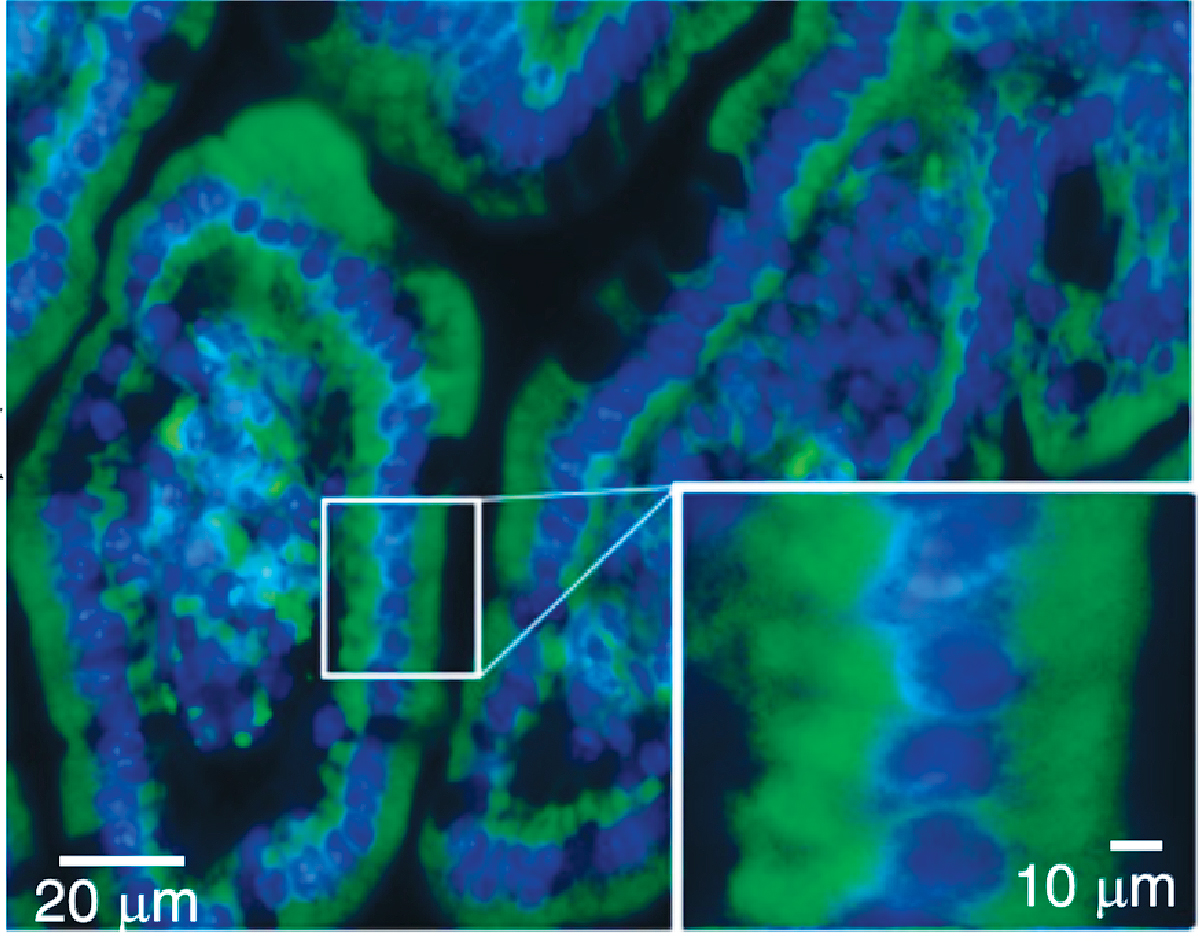
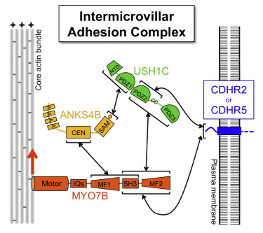
Adhesion protein optimizes border
Feb. 14, 2019, 10:45 AM by Leigh MacMillan (iStock) The epithelial cells that line the intestines build a specialized cell surface — the “brush border” — that processes and absorbs nutrients, and defends against pathogens. The brush border consists of thousands of finger-like membrane protrusions (microvilli) on each… Read MoreFeb. 15, 2019
-

Elongating Microvilli in the Intestine
Elongating Microvilli in the Intestine Enterocyte showing Brush border and unstirred layer Author: Boumphreyfr Enterocytes, the epithelial cells lining the lumen of the intestine, develop a dense lawn of microvilli (MV) on their apical surface as they differentiate from stem cells in the intestinal crypt. We know that MV… Read MoreOct. 10, 2018
-
Sulfate-Iron Link to Anemia
Sulfate-Iron Link to Anemia Sulfation (the addition of a sulfate group to a molecule) is an important biochemical process that aids in the detoxification of xenobiotic compounds and plays a role in the biosynthesis of a variety of molecules. In mammalian cells, sulfation requires PAPS (3´-phosphoadenosine 5´-phosphosulfate), which donates… Read MoreMar. 27, 2018
-

Cell skeleton and the brush border
The epithelial cells lining organs like the intestines and kidneys build a special surface called the “brush border,” which consists of a dense array of finger-like protrusions. Irina Kaverina, PhD, Matthew Tyska, PhD, and colleagues in Argentina explored the role of microtubules — part of the cellular “skeleton” — in building the… Read MoreFeb. 1, 2018
-
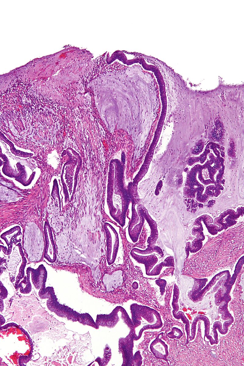
Drivers of Intestinal Tumorigenesis
Drivers of Intestinal Tumorigenesis A hallmark of all epithelia is the presence of adherens junctions that connect adjacent cells to each other. The junctions are formed through the interaction of the extracellular domains of E-cadherin on the neighboring cells. In turn, the intracellular domain of E-cadherin forms a complex… Read MoreDec. 6, 2017
-
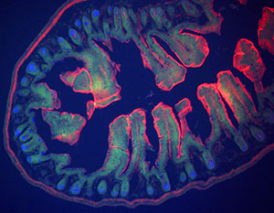
Tracing Cell Origins in the Gut
Tracing Cell Origins in the Gut The organs of multicellular animals comprise highly organized aggregates of many cell types, each of which has differentiated from a multi-potent stem cell. Although we have learned much about the process of differentiation and organogenesis through studies of tissues such as bone marrow,… Read MoreDec. 5, 2017
-

Gut response to fluid flow
Flow of fluids through the gut, such as milk from an infant’s diet, generates a shear stress on cells lining the intestine. Ken Lau, Ph.D., and colleagues have demonstrated that microvilli – finger-like membrane protrusions – are capable of sensing shear forces and subsequently drive an intracellular response called autophagy. Read MoreOct. 26, 2017
-
Key to Brush Border Assembly in the Intestine
A primary function of the lining surface of the intestine is to absorb nutrients. The epithelial cells that form this surface are notable for the presence of a brush border composed of microvilli, tiny plasma membrane projections that markedly increase the surface area through which absorption can take place. Read MoreDec. 2, 2016