Biopsies are clinical tools commonly used to diagnose a variety of diseases or to monitor tissue for abnormal growth or even rejection of a transplant. During biopsies, tissue samples are removed from the body so they can be examined more closely, but depending on the type of tissue that’s needed, the procedure can be rather invasive.
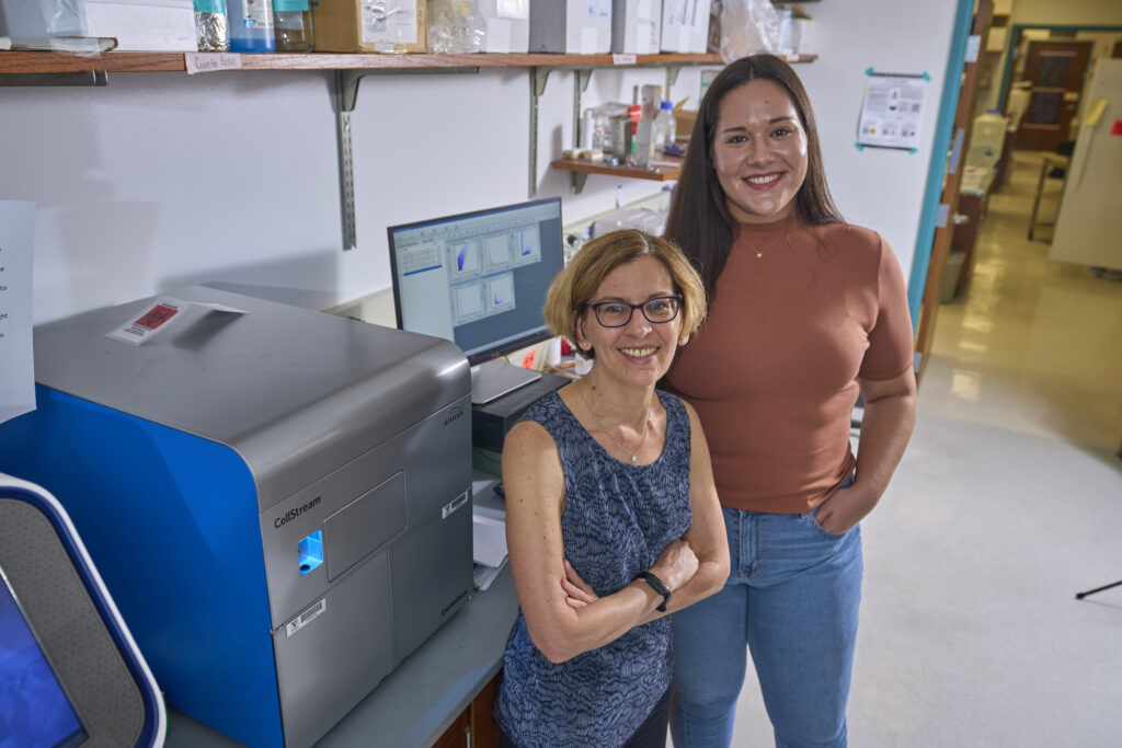
Researchers from the School of Medicine Basic Sciences recently developed an analytical tool that could lead to the use of “liquid biopsies” as a substitute for traditional biopsies for certain patients or diseases. The tool, called EV Fingerprinting, was the culmination of the dissertation work of Ariana von Lersner, a former graduate student and current postdoctoral scholar in the laboratory of Alissa Weaver, Cornelius Vanderbilt Professor of Cell and Developmental Biology.

The ”EV” in EV Fingerprinting stands for extracellular vesicles, which are membrane-bound particles that contain biologically active cargo and that contribute to cell-cell communication in health and disease. Although EVs have been observed since at least the 1980s, their origin and purpose have not been clearly defined. The last two decades have seen research into EVs skyrocket, and EVs have now been found to have roles in endocrine processes, immune responses, and even cancer progression in a variety of species, including humans.
The term “EV” encompasses vesicles of various sizes and cargos, each likely tailored to different functions. Changes in the heterogeneity of EVs in an organism can reflect changes in biological state—for example, a cancer state vs. a normal, nondisease state—which can serve as a clinically informative biomarker.
“Fingerprinting allows you to characterize EVs with minimal sample preparation in a high-throughput manner, and allows you to better classify the types of vesicles in the sample,” von Lersner said.
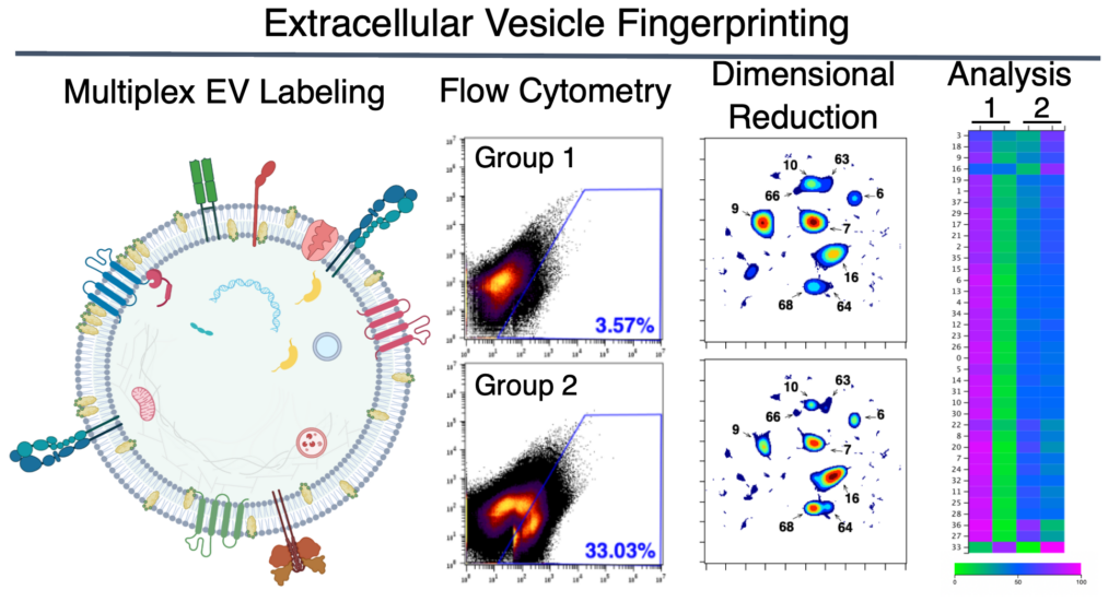
The technique involves isolating EVs from the rest of the cellular content in a sample, labeling them with a fluorescent lipophilic dye that intercalates into the EVs’ lipid bilayer, and running them through a flow cytometer, an instrument that shoots a laser at a sample and collects information about how the light is refracted or emitted. The collected information is compiled into a “fingerprint” that can be used to perform quantitative analyses of distinct EV populations and determine how they are altered by experimental manipulation, molecular perturbation, or disease state.

EV Fingerprinting constitutes an unprecedented advancement toward the characterization of EVs because it can analyze the composition of the lipid bilayers of the EVs in a sample and breaks the sample down into individual EV populations, which previous bulk analysis methods could not do. Using the composition of the lipid bilayers to separate EV populations is a novel approach that capitalizes on an EV characteristic that had been previously overlooked by the field.
The work was completed thanks to the contributions of Vanderbilt collaborators from the departments of cell and developmental biology, chemical and biomolecular engineering, and pathology, microbiology and immunology and the Center for EV Research, and external collaborators from the Cedars-Sinai Medical Center and Genentech. The Center for EV Research, established in 2021, is managed under Weaver’s direction and provides shared instrumentation and training for EV work, fosters interdepartmental discussion and collaboration through monthly seminars and annual retreats, and provides members with funds to conduct or share EV-related work at conferences.
“EV Fingerprinting is furthering the development of liquid biopsies in which the EVs can be used as biomarkers for diseases such as cancers or neurological disorders,” von Lersner said.
If you find yourself needing a biopsy one day and can forgo the traditional kind in favor of a simple blood draw, you may have these researchers to thank.
Go deeper
The paper “Multiparametric Single-Vesicle Flow Cytometry Resolves Extracellular Vesicle Heterogeneity and Reveals Selective Regulation of Biogenesis and Cargo Distribution” was published in ACS Nano in April 2024.
Funding
This research was funded by the National Institutes of Health and the National Science Foundation.