The neural crest regulates beta cell proliferation and maturation
Received 30 June 2010;
revised 27 October 2010;
accepted 5 November 2010.
Available online 14 November 2010.
Abstract
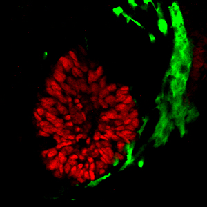
Interactions between cells from the ectoderm and mesoderm influence development of the endodermally-derived pancreas. While much is known about how mesoderm regulates pancreatic development, relatively little is understood about how and when the ectodermally-derived neural crest regulates pancreatic development and specifically, beta cell maturation. A previous study demonstrated that signals from the neural crest regulate beta cell proliferation and ultimately, beta cell mass. Here, we expand on that work to describe timing of neural crest arrival at the developing pancreatic bud and extend our knowledge of the non-cell autonomous role for neural crest derivatives in the process of beta cell maturation. We demonstrated that murine neural crest entered the pancreatic mesenchyme between the 26 and 27 somite stages (approximately 10.0 dpc) and became intermingled with pancreatic progenitors as the epithelium branched into the surrounding mesenchyme. Using a neural crest-specific deletion of the Forkhead transcription factor Foxd3, we ablated neural crest cells that migrate to the pancreatic primordium. Consistent with previous data, in the absence of Foxd3, and therefore the absence of neural crest cells, proliferation of insulin-expressing cells and insulin-positive area are increased. Analysis of endocrine cell gene expression in the absence of neural crest demonstrated that, although the number of insulin-expressing cells was increased, beta cell maturation was significantly impaired. Decreased MafA and Pdx1 expression illustrated the defect in beta cell maturation; we discovered that without neural crest, there was a reduction in the percentage of insulin-positive cells that co-expressed Glut2 and Pdx1 compared to controls. In addition, transmission electron microscopy analyses revealed decreased numbers of characteristic insulin granules and the presence of abnormal granules in insulin-expressing cells from mutant embryos. Together, these data demonstrate that the neural crest is a critical regulator of beta cell development on two levels: by negatively regulating beta cell proliferation and by promoting beta cell maturation.
Research Highlights
► Neural crest cells first enter the pancreatic primordium at the 27 somite stage.
► Neural crest reaches the dorsal pancreas prior to the ventral pancreas.
► In the absence of neural crest, insulin-expressing area is increased.
► Neural crest is required for beta cell maturation.
► Neural crest derivatives directly contact beta and alpha cells by 15.5 dpc.