2018 Cell Dynamics Image Winners
 |  |  |
| +wntcomposite3 | +wntcomposite2 | -wntcomposit |
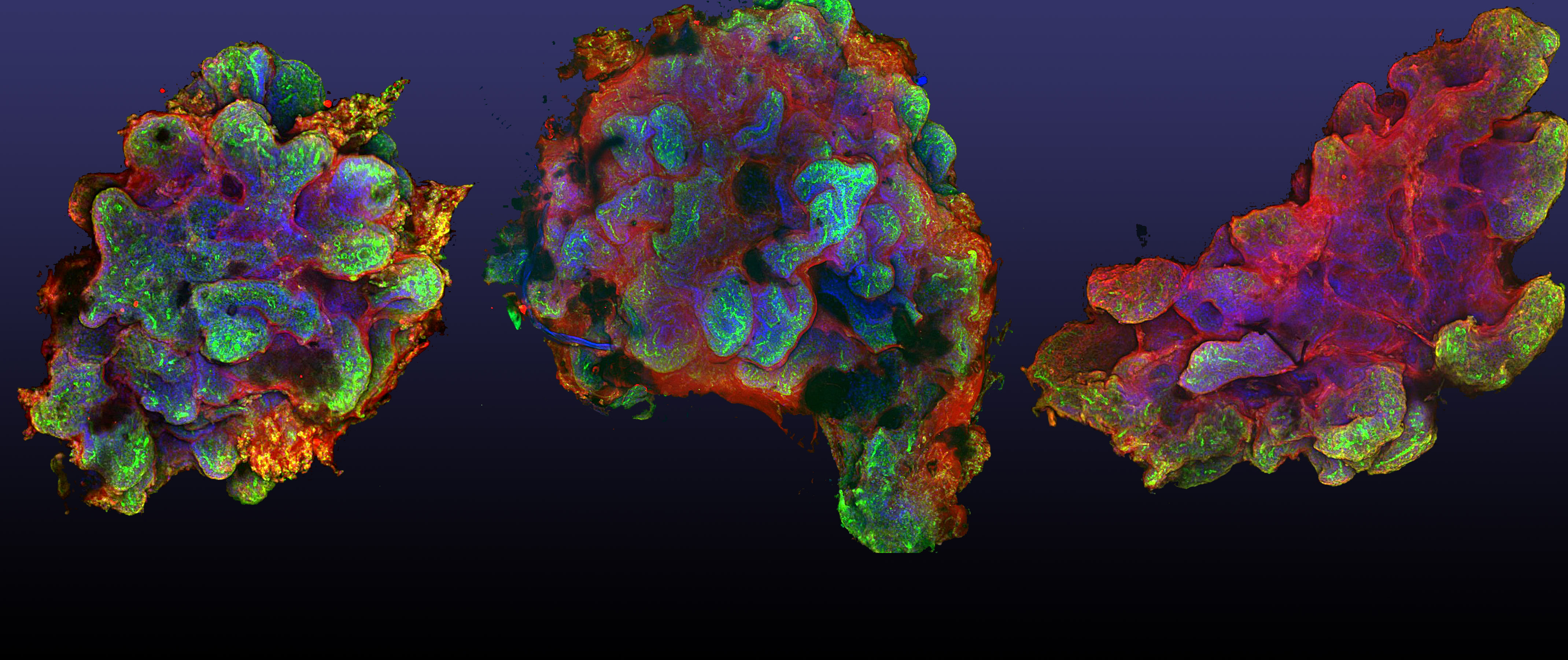 |
| Images combined to show Tryptic image |
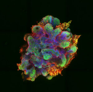
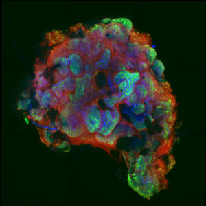
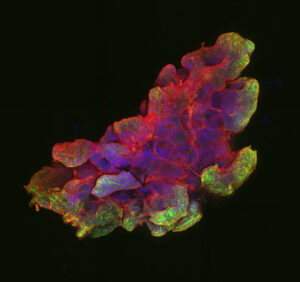
Human intestinal organoids generated from IPS cells. Beta-catenin is (red), actin (green), and DNA (blue) [Images by Leah Sawyer (Lee Lab)]
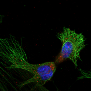
The Great Divide. Previous data from our lab indicated that an E3 ligase, Cullin 9 (Cul9), localizes to mitotic spindles in human pluripotent stem cells. Our data shows that Cul9 (Red) also localizes to the mitotic spindle (Green), in the human fibroblasts (HdFns). Image was taken at 63x using AiryScan with help from Natalya Ortolano. [(Image by Mary Chalkley (Gama Lab)]
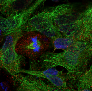
Tubulin’s guide to the galaxy. Previous data from our lab indicated that an E3 ubiquitin ligase localized to the mitotic spindle in human pluripotent stem cells. Staining for alpha-tubulin (green) and the E3 ubiquitin ligase (red) in neural stem cells shows a different, seemingly non-specific localization. [(Image by Natalya Ortolano (Gama Lab) / Staining by Mary Chalkley (Gama Lab)]