Discoveries
-
ATAC-Me attacks knowledge gap in genetics research
https://cdn.vanderbilt.edu/t2-main/medschool-prd/wp-content/uploads/sites/101/2020/03/Hodges.mp4 By Hillary Layden Most human cells contain roughly 6.5 feet of DNA, which must be tightly compacted to fit within the nucleus. Cells compact DNA by wrapping it around proteins called histones, forming a DNA-protein complex called chromatin. “Closed” chromatin is tightly compacted and cannot interact with… Read MoreMar. 17, 2020
-

Pancreatic islet cells distinct in mice and humans
By Cassandra Awgulewitsch https://cdn.vanderbilt.edu/t2-main/medschool-prd/wp-content/uploads/sites/101/2020/02/SteinDiabetes.mp4 Researchers in the lab of Roland Stein (Molecular Physiology & Biophysics), along with collaborators at UCSF, NC State, and UPenn, have shown distinct changes in human pancreatic islet cells throughout the course of life and the progression of type 2 diabetes. They also discovered… Read MoreFeb. 19, 2020
-

The tale of the targeted mouse
By Sarah Glass https://cdn.vanderbilt.edu/t2-main/medschool-prd/wp-content/uploads/sites/101/2020/02/Coffey_Updated.mp4 3D illustration of colorectal cancer. Kateryna_Kon, stock.adobe.com. Researchers from the labs of Robert Coffey (Medicine) and Jacob Houghton (Radiology and Radiological Sciences) report in Gastroenterology the identification of two human antibodies, P1X and P2X, that can neutralize EGFR in mice. EGFR,… Read MoreFeb. 13, 2020
-
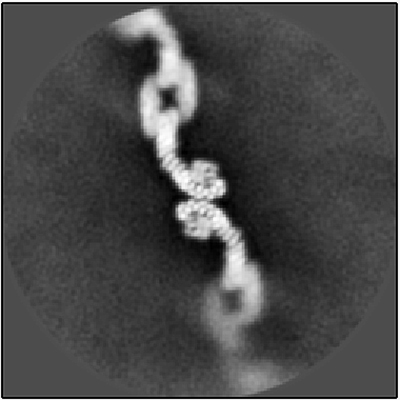
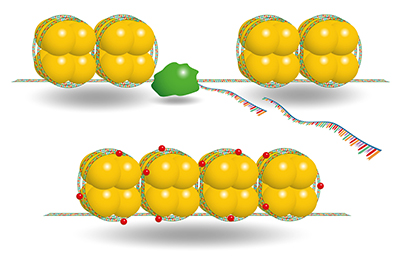
A master cellular conductor
https://cdn.vanderbilt.edu/t2-main/medschool-prd/wp-content/uploads/sites/101/2020/02/Jackson_updated.2-1.mp4 This article was submitted by the senior author of the featured paper, Lauren P. Jackson. Cryo-EM image of retromer chain assembly. Courtesy of Amy K. Kendall Human cells contain a “FedEx system” to ensure that important protein and fatty lipid cargo molecules are delivered to the right… Read MoreFeb. 12, 2020
-

Setting up DNA repair
By Alexandria Oviatt DNA repair pathways such as NER have the integral role of protecting us from potentially damaging mutations. Defects in these mechanisms can lead to diseases such as XP or cancers. (Gernot Krautberger, stock.adobe.com) A recent Nucleic Acids Research paper from the lab of Walter… Read MoreFeb. 6, 2020
-

Receptor modulators chart new courses out of depression
By Amanda N. Johnson “Major depression is one of the most common mental disorders in the U.S. According to the National Institute of Mental Health, approximately 7% (17.3 million) of American adults had at least one major depressive episode in 2017.” (Tadamichi, stock.adobe.com) Existing drug treatments relieve mental illness for… Read MoreFeb. 4, 2020
-

Breaking up MYC-WDR5 to counter cancers
By Suneethi Sivakumaran C-MYC, a variant of MYC, and MAX bound to DNA. (Molekuul.be, stock.adobe.com) Cancers are complex and diverse in nature, assailing the human body through different mechanisms. Cancer cells outsmart normal cells through myriad mechanisms, including sustained proliferation, insensitivity to growth suppressors, and resistance to cell… Read MoreFeb. 4, 2020
-

Targeting NA to protect against lethal avian flu infection
By Sohini Roy New research can lead to improved vaccines against the flu, including strains such as H7N9 and antiviral-resistant strains. Image by Heather Hazzan, SELF Magazine. Published under a CC BY 2.0 license. Obtained from Flickr. Asian lineage avian influenza virus (H7N9) is… Read MoreJan. 22, 2020
-

Of mice and tailgaters: Identifying neural circuitry involved in binge drinking
By Deborah Roby A mouse drinking from a water dispenser. Published under a CC0 1.0 license. Researchers at the Vanderbilt Center for Addiction Research, along with collaborators at MIT and Salk Institute, have determined a neurological pathway that may be used to determine a… Read MoreDec. 19, 2019
-

Fight or flight – Flexibly
By Julia Thompson Artist’s rendering of a mental health concept, by Quince Media. Image reproduced under a CC BY 4.0 license. Fear is a crucial emotion for human survival. Without the ability to experience fear in response to possible threats in the environment, it is all too easy… Read MoreDec. 19, 2019